Anatomy and Cell Biology 3309 Lecture Notes - Lecture 15: Periodontal Fiber, Loose Connective Tissue, Neural Crest

22 May 2018
School
Department
Professor

Histology 3309
Teeth
Learning Objectives: Teeth
1. Prepare a labeled diagram of the adult tooth in sagittal section
2. State the main physical property of dentin & describe its organic & inorganic constituents.
3. Describe the location & structure of odontoblasts & their role in producing dentin.
4. State the main physical property of enamel & describe its organic & inorganic constituents.
5. Name the location & structure of ameloblasts & their role in producing enamel.
6. Explain the location, histology and the functions of the periodontal ligament.
7. State the components of tooth pulp.
8. Explain the location and composition of cementum.
9. Describe the development of the tooth, including the formation of dentin, enamel, and
cementum.
Dentition
- The set of teeth we have is called the dentition
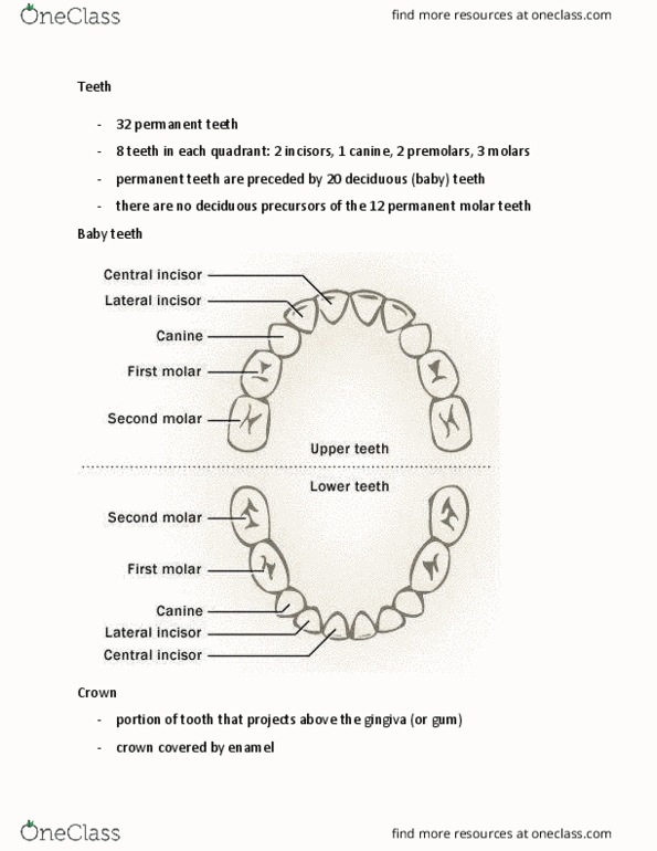
- 32 permanent teeth (ideally)
- 8 teeth in each quadrant:
o 2 incisors
o 1 canine
o 2 premolars
o 3 molars
- the molars come in fairly late while the incisors, canine and premolars come in
as baby teeth and we lose them and they get replaced with permanent teeth
- at the back we have wisdom teeth which come in very late – sometimes there is
not enough room and so they grow crooked and so they need to be removed
- permanent teeth are preceded by 20 deciduous (baby) teeth
- no deciduous precursors of the 12 permanent molars
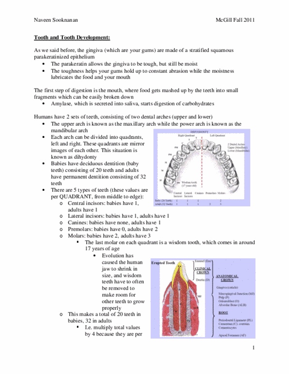
- this image divides the tooth into diff parts
- the body of the tooth is made of a material called dentin (main calcified/mineralized material of
the tooth)
- however, it’s a living material there are cellular components inside
- there are also other mineralized structures as well – enamel
- enamel covers the dentin on the outside of the tooth
- enamel is the hardest material in the body as it is highly mineralized
- we also have another mineralized material called cementum
- cementum covers the part of the tooth that you don’t see – it anchors the bone to the alveolar
bone
- the tooth is divided into 2 regions:
o root
▪ the dentin part is covered by cementum
o crown
▪ the dentin part is covered by enamel
▪ you don’t see all parts of the crown
▪ the crown is divided into…
• anatomical crown
o identifies where the cementum starts
• clinical crown
o part of the tooth that projects from the gingival socket
(indentation of your epithelium/epidermis)
find more resources at oneclass.com
find more resources at oneclass.com

o this is where food can get stuck and we can get tooth decay
- note: tooth is hollow
- it has a chamber filled with loose connective tissue called pulp
o there are lots of nerves and blood vessels that come in here to nourish the tooth
o the cells that produce dentin aligning the pulp chamber and they need nutrition and
oxygen and waste removal etc
- the tooth also needs to be anchored relatively firmly to the alveolar bone
o so there is a connection bw the cementum and the alveolar bone called the periodontal
ligament (dense connective tissue)
o it is important that this ligament is able to regenerate
o it leaves a little bit of room for movement (so your teeth wiggle to a certain extent)
o if bacteria come into that region and start eating at your periodontal ligament, you could
lose your teeth
o this is also the area that is touched to straighten the tooth
1. Bud Stage:
- early on the epithelium that lines the oral cavity starts to form growth inot the underlying
mesenchyme (the embryonic loose connective tissue(
- what induces the formation of this tooth bud is the accumulate of neural crest cells that migrate
underneath the oral epithelium and locally induce the epithelium
to grow into the mesenchyme
- the neural crest cells have an important function in forming part
of the tooth
- neuroectodermal cells induce the overlying
epithelial cells to proliferate and form an
invaginating tooth bud
find more resources at oneclass.com
find more resources at oneclass.com

2. Cap Stage
- the tooth bud forms an upside down up (indention)
- in response to signals that come from the neural crest and the mesenchyme
- the neural crest cells that were underneath the tooth bud accumulate and form a layer
- the remaining mesenchyme forms a round structure of cells called the dental sac
- the neural crest and the dental sac forms diff parts of the tooth
- the epithelial tooth bud forms a cup-like structure under the influence of cytokines secreted by
cells in the mesenchyme
3. Late Cap Stage
- the dental papilla (mesenchyme) expands and the neural crest cells form a layer of epithelial cells
underneath the oral epithelium that came in
- now they are called preondontoblasts
- Cells that make dentin and are derived from neural crest
- The overlying epithelium from the mouth cavity develops into a layer of cells called ameloblasts
(early on they are called preameloblasts) - So we called them the enemal epithelium – these cells
produce enamel
- Those cells at this stage are next to one another and there is no enamel or dentin formed yet
- the epithelial tooth bud is lined by an outer and inner enamel epithelium
- the inner enamel epithelium differentiates into a single layer of ameloblasts
- odontoblasts develop from the cranial neural crest in the dental papilla
find more resources at oneclass.com
find more resources at oneclass.com
Document Summary
The set of teeth we have is called the dentition. Permanent teeth are preceded by 20 deciduous (baby) teeth. No deciduous precursors of the 12 permanent molars this image divides the tooth into diff parts the body of the tooth is made of a material called dentin (main calcified/mineralized material of the tooth) Cells that make dentin and are derived from neural crest. The overlying epithelium from the mouth cavity develops into a layer of cells called ameloblasts (early on they are called preameloblasts) - so we called them the enemal epithelium these cells produce enamel. Purpose of this hard material is : protect the dentin, provide a surface for the periodontal ligament to make a connection to the alveolar bone are formed by odontoblasts that are sitting on the outside of cementum vessel. 45-50% organic material (mainly type 1 collagen) Anchor periodontal ligament into cementum and alveolar bone.