Anatomy and Cell Biology 3309 Lecture Notes - Lecture 12: Neural Groove, Neural Tube, Neural Fold

19 Aug 2019
School
Department
Professor

ACB 3309 Lecture 12 (Nervous Tissue and its Organization)
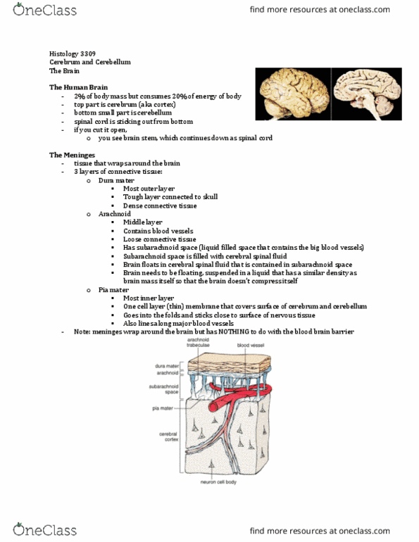
CNS – Cerebrum and Cerebellum
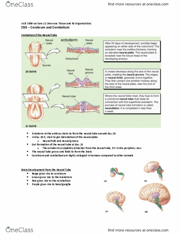
Formation of the Neural Tube
Ectoderm in the embryo starts to form the neural tube around day 20
At day 20.5, start to get indentation of the neural plate
o Neural fold and neural groove
Get formation of the neural tube at day 21
o The ectoderm completely detaches from the neural tube, it’s in the periphery now
The neural tube grows and folds to form the brain
Cerebrum and cerebellum are highly enlarged in humans compared to other animals
Brain Development from the Neural Tube
Beige gives rise to cerebrum
Green gives rise to the brainstem
Red gives rise to the cerebellum
Purple gives rise to basal ganglia

• Cerebral & cerebellar cortex are highly folded to package a huge amount
of neurons into the cranial cavity
• Cerebral cortex: bumps are called gyri, valleys between are called sulci
o Important for sensory processes, language, movement,
• Cerebellar cortex: parallel ridges are called folia
o Motor control and coordination
o Cross section looks like a leaf
• More SA the more synapses that can occur, so more efficient
o These folds are increasing SA, to increase the area for the
synapses to occur
• Brainstem is continuous w the spinal cord
White and Gray Matter
Gray matter
o Nerve cell bodies
o Neuropil – dendrites, glial cells, synapses
and unmyelinated axons of interneurons
White matter
o Myelinated axons & associated glial cells
(e.g. oligodendrocytes and Schwann cells)
Right = gray matter
o motor neuron:
▪ massive cell body
▪ prominent nucleiolus and pale nucleus
(w tons of euchromatin)
▪ dark stained areas = nissl bodies (free
ribosomes w/in the perinuclear
cytoplasm)
o Motor neuron is MUCH bigger than glial cells (G)
o Neuropil = connective tissue of nervous system
Left = white matter
o Where myelinated axons are gonna pass through
▪ White = myelin
▪ Dark stained = axon
o Need special stain to stain lipid
o In H&E, stain is washed away so looks white/pale
o So white matter is more palely stained