Anatomy and Cell Biology 3319 Lecture Notes - Lecture 7: Interthalamic Adhesion, List Of Thalamic Nuclei, Intralaminar Nuclei Of Thalamus

29 Sep 2016
School
Department
Professor
Document Summary
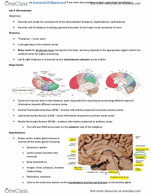
Subthalamus (includes subthalamic nucleus from basal ganglia) Two halves of thalamus on either side of midline mirrors images. Third ventricle lies right at midline beneath basal ganglia. Thalamus is on either side of that third ventricle. Fornix is fibril track connecting parts of temporal lobe (hippocampus to thalamus?) Medial and posterior to basal ganglia overlaps to some extent but more medial. When you slice into thalamus you have series of nuclei. The diencephalon includes the thalamus, hypothalamus, epithalamus (pineal) and the subthalamus. The thalamus is the largest component of the diencephalon and is located medial to the internal capsule at the midline on either side of the third ventricle. The thalamus is ventral to fornix, a large band of fibres involved in limbic connections. The right and left halves of the thalamus are connected by the interthalamic adhesion that extends through the third ventricle. The pineal gland is located posterior to the thalamus.