Anatomy and Cell Biology 3319 Lecture Notes - Lecture 20: Ependyma, Occipital Lobe, Neural Tube

1 Nov 2016
School
Department
Professor
Document Summary
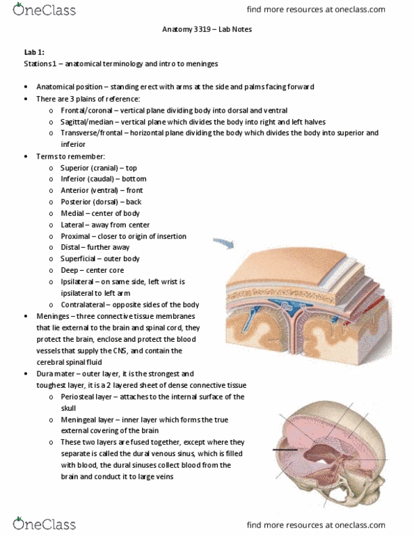
Connective tissue coverings of brain and spinal cord: dura mater, arachnoid mater, pia mater. Two layers of dense fibrous connective tissue. Periosteal layer close to bone (skull calaveras) Meningeal layer closer to brain and continues down to spinal cord and forms dural sac. Middle meningeal layer that spans over the surface of the gyri. Filaments (green lines) anchored to pia mater giving spider web appearance. Through arachnoid mater is where you"ll have csf. Pia mater (tender mother) thin red line. Firmly adhered to the surface of the brain, spinal cord, and origin of cranial nerves invaginates into sulci. Space b/w arachnoid and pia (small green lines filaments) Potential or true spaces where fluid can accumulate. Potential spaces can become a space if you have fluid accumulation (ex trauma: epidural space (above dura mater, subdural space (under dura mater) Accumulation of blood between the periosteal dura mater and the skull (puts pressure on brain so want to drain out blood.