Anatomy and Cell Biology 3319 Lecture 33: Coronary and Pulmonary Circulation

13 Feb 2017
School
Department
Professor

Document Summary
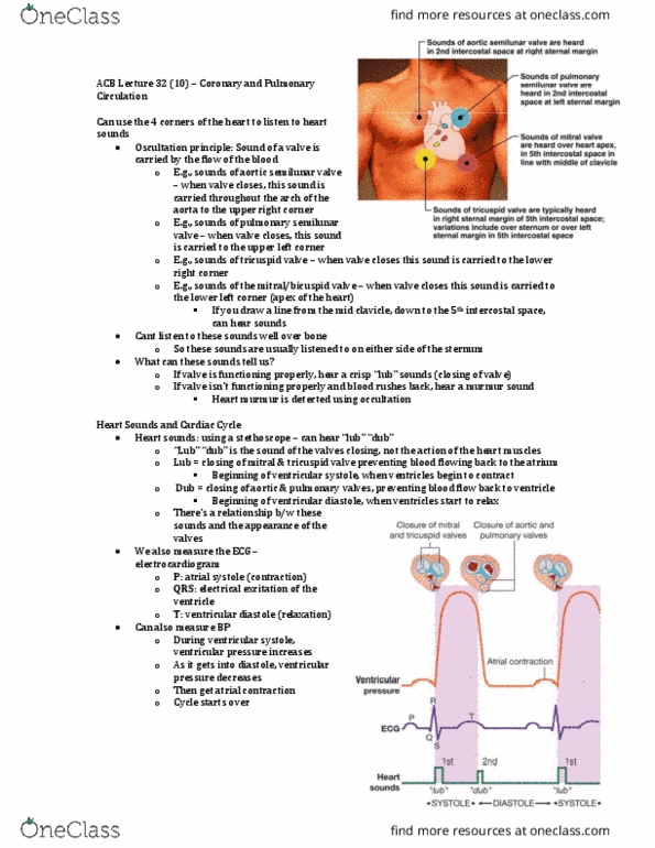
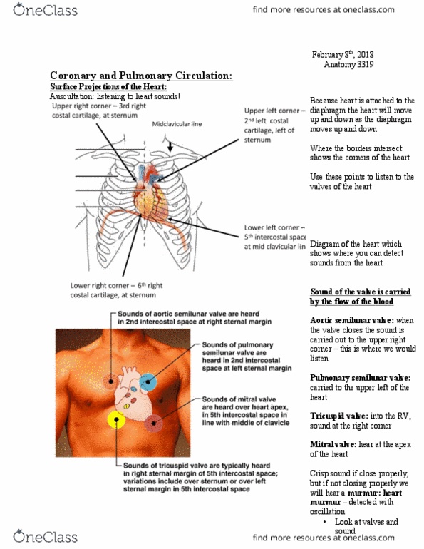
Des(cid:272)ri(cid:271)e : principles of auscultation, congenital defects of the heart, conductile system of the heart, coronary circulation of the heart, pulmonary circuit, 3 major branches coming off the arch of the aorta. Re(cid:272)all the 4 (cid:271)orders & 4 (cid:272)or(cid:374)ers of the heart fro(cid:373) the (cid:862) urfa(cid:272)e proje(cid:272)tio(cid:374)s of the heart(cid:863) slide: upper left corner: 2nd left costal cartilage, left of sternum, upper right corner: 3rd right costal cartilage, at sternum. Lower left corner: 5th intercostal space, at midclavicular. Lower right corner: 6th costal cartilage, at sternum. A sethoscope must be strategeically placed on the chest wall to listen to the individuall sounds of the 4 valves. The sound of a valve is carried by the flow of blood: therefore, the semilunar valves will have their heart sound at the top of the heart. Aortic semilunar valve: upper left corner 2nd intercostal space, at right sternal margin.