Anatomy and Cell Biology 3319 Lecture Notes - Lecture 19: Medial Pterygoid Muscle, Lateral Pterygoid Muscle, Myocyte

1 May 2018
School
Department
Professor

Lecture 019: Introduction to the Muscular System: Muscles of the Head
Objectives
● Understand basic muscle structure and terminology
● Describe the attachments, actions and innervation of the 12 major muscles of facial
expressions
● Describe the attachments (origin/insertion), action and innervation of the 4 muscles of
mastication
○ Masseter, temporalis, lateral, medial pterygoid
● Describe the extrinsic (genioglossus, hyoglossus, styloglossus) muscles of the tongue
● Identify the cranial nerves which supply these muscle groups
● Predict the consequences of nerve damage to any of these muscle groups
● Describe a quick method to assess the function of theses nerves
Introduction to the Muscular System
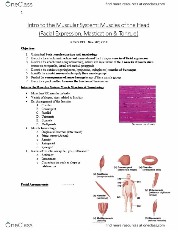
● More than 700 muscles in the body, variety of shapes and sizes related to function
○ Arrangements of the fascicles
■ Circular
● Arranged in a circle
● Sphincters
■ Convergenet
● Origin of the muscle is in a fan shape
● All of the fibers converge into one point
○ Since the fibers are pointing to different locations it allows
for multiple actions
■ Parallel
● Long muscle fiber
● Only contact in one direction, thus only one main action
● But it can’t shorted too much - not very powerful
■ Unipenate
● “Feather like”
● Tendon down the middle, fibers coming off it
● One side
■ Bipenate
● Fibers coming out of both side of the tendon
■ Multipennate
● Multiple bipennate muscles put together
● Strong and big (deltoid)
■ Fusiform
● Biceps
● Tapered at the ends, thick in the middle
● Muscle terminology
○ Origin and insertion (attachment)
■ Origin: where it begins
find more resources at oneclass.com
find more resources at oneclass.com

■ Insertion: where the tendon attaches to the bone
○ Prime mover
■ Major action
■ Agonist
○ Antagonist
■ Muscle which opposes the action (agonist)
○ Synergist
■ Muscle that assists with the main action
○ Need a good balance between the agonist and antagonist to have good actions
● Names of muscles always tell you something about
○ Action it performs
○ Location
○ Characteristic such as shape (ex. deltoid) or relative size
Muscles of Facial Expression
● Originate on a bone
● But insert on skin or interminles with other muscles
● Sphinctor/dilators around orfices of the faces
○ Sphincter:
■ Arranged circularly
■ Around the mouth and eyes
○ Dilators
■ More radially arranged (pull the orifices open)
Muscles of Facial Expression
● Know what bone the come from, and skin it interminges with
1. Orbicularis oculi
● Orbicularis: circular muscle
● Oculi: eye
● Origin: medial palpebral ligament
● Inserts: skin around eye
● Contraction of the palpebral: close eye softly (blinking)
○ Located in the eyelid
● Contraction of the orbital: closes eyes tightly (squinting)
○ Surrounds the orbit
2. Corrugator supercilii
● Origin: medial end superciliary arch
● Inserts: skin superior to the middle of the supraorbital margin
● Contractions: draw eyebrows medially and inferiorly, creates wrinkles above the nose
○ A frown
3. Orbicularis oris
find more resources at oneclass.com
find more resources at oneclass.com
Document Summary
Lecture 019: introduction to the muscular system: muscles of the head. Describe the attachments, actions and innervation of the 12 major muscles of facial expressions. Describe the attachments (origin/insertion), action and innervation of the 4 muscles of mastication. Identify the cranial nerves which supply these muscle groups. Describe the extrinsic (genioglossus, hyoglossus, styloglossus) muscles of the tongue. Predict the consequences of nerve damage to any of these muscle groups. Describe a quick method to assess the function of theses nerves. More than 700 muscles in the body, variety of shapes and sizes related to function. Origin of the muscle is in a fan shape. All of the fibers converge into one point. Since the fibers are pointing to different locations it allows. Only contact in one direction, thus only one main action. But it can"t shorted too much - not very powerful. Tendon down the middle, fibers coming off it. Fibers coming out of both side of the tendon.