Anatomy and Cell Biology 3319 Lecture Notes - Lecture 13: Primary Olfactory Cortex, Optic Nerve, Olfactory Bulb

1 May 2018
School
Department
Professor

Lecture 013: Cranial Nerves
Objectives
● Name the 12 pairs of cranial nerves
● List the individual functions of each cranial nerve
● Diagnose cranial nerve damage based on symptoms
Cranial Nerve Overview
● 12 pairs of cranial nerves
● Emerge off the VENTRAL surface of the brain
● Numbered 1-12 from the rostral to caudal (I - XII)
● All innervate the head and neck (except Vagus)
● 2 emerge from the forebrain
● 10 emerge from brainstem
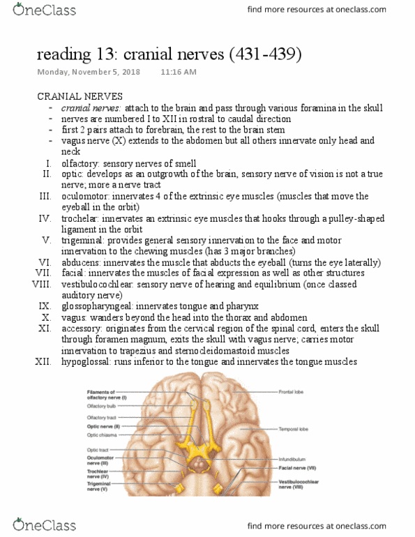
Cranial Nerve
I: OLFACTORY NERVE/BULB
● Olfactory receptor cells -> olfactory bulb -> olfactory tract -> 1o olfactory cortex
○ Filaments in the nasal cavity line the nasal mucosa
○ Odorants come into nose and activate the filaments which sends AP to the
olfactory receptor cells
○ Receptor cells synapses in the olfactory bulb, travels via the olfactory tract to the
primary olfactory cortex (temporal lobe)
● Function: smell (olfaction)
II: OPTIC NERVE
● Retina -> optic nerve -> optic chiasm -> optic tract -> LGN -> 1o visual cortex
○ Light hits retina
■ Retina is divided into 2 halfs
● Nasal retinal: midline
● Temporal retinal
:lateral/peripheral
● Nasal retinal information
crosses over the the
optic chiasm
■ However, each retina will have
a visual field that OVERLAPS
with the other one
● Binocular visual field (area where both visual fields overlap)
○ Temporal retina
○ Information of the visual fields crosses at the optic chiasma
■ After (in the optic tract) visual information will be similar (all right or left
visual information)
○ Lateral Geniculate Nucleus (LGN)
find more resources at oneclass.com
find more resources at oneclass.com

○ Information send to the 1o visual cortex for conscious perception
○ Damage of cranial nerve II:
■ At optic nerve: loss of some ipsilateral peripheral visual field
■ At optic chiasm: bilaterally (BOTH) peripheral
field loss
● Only binocular visual field left
■ At optic tract: loss of ENTIRE contralateral
visual field
● Function: sight (vision)
● Pituitary gland sits posterior to optic chiasm
Pre-Test
● Symptoms
○ Limited to binocular vision
○ Bilateral peripheral blindness (bitemporal hemianopia)
● What cranial nerve is involved?
○ Cranial nerve II (at the optic chiasm)
● What is the diagnoses?
○ Pituitary tumor (enlargement of the pituitary gland
III: OCULOMOTOR NERVE
● Arise from the midbrain and travel into the eye-socket
● Motor (innervates)
○ Medial rectus, Inferior rectus,
Superior rectus, Inferior oblique
■ Inserts onto the eyeball itself
○ Levator palpebrae superioris
■ Inserts onto the eyelid (open
and close eyelid)
● Parasympathetic
○ Synapse in the ciliary ganglion and
enter eye
○ Innervates the sphincter papillae in
the iris
■ Allows for pupillary
constriction (controls how
much light is let in)
○ Innervate the ciliary muscles
■ Controls the thickness of the lens (allows it to thicken and focus at closer
distances)
IV: TROCHLEAR NERVE
● Dorsal brainstem -> superior orbital fissure -> eye
socket
find more resources at oneclass.com
find more resources at oneclass.com
Document Summary
Name the 12 pairs of cranial nerves. List the individual functions of each cranial nerve. Diagnose cranial nerve damage based on symptoms. Emerge off the ventral surface of the brain. Numbered 1-12 from the rostral to caudal (i - xii) All innervate the head and neck (except vagus) Olfactory receptor cells -> olfactory bulb -> olfactory tract -> 1o olfactory cortex. Filaments in the nasal cavity line the nasal mucosa. Odorants come into nose and activate the filaments which sends ap to the olfactory receptor cells. Receptor cells synapses in the olfactory bulb, travels via the olfactory tract to the primary olfactory cortex (temporal lobe) Nasal retinal information crosses over the the optic chiasm. However, each retina will have a visual field that overlaps with the other one. Binocular visual field (area where both visual fields overlap) Information of the visual fields crosses at the optic chiasma.