Anatomy and Cell Biology 3319 Lecture Notes - Lecture 31: Middle Cardiac Vein, Common Carotid Artery, Inferior Vena Cava

1 May 2018
School
Department
Professor

Lecture 031: Heart Structure and Function
Objectives
● Describe the location and surface anatomy of the heart.
● Describe anatomical features of chambers and valves of the heart
● Indicate where on the chest wall each valve can be heard
● Describe the conducting system of the heart
● Compare fetal and adult circulation
Circulation Overview
● Pulmonary Circulation
● Coronary Circulation
○ Supplies the heart
○ If compromised can lead to heart attacks
● Systemic Circulation
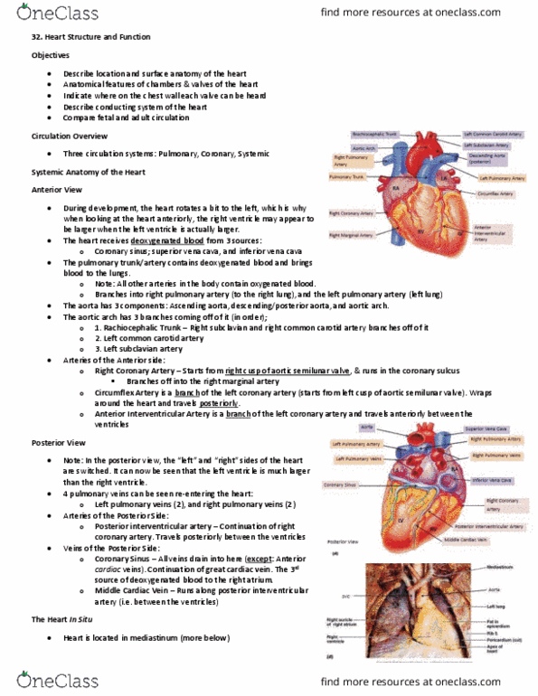
Surface Anatomy of the Heart
● 4 chambers
○ RA, LA, RV, LV
○ The LA will be slightly posterior due to the heart rotating during development
● Great vessels
○ Superior and Inferior Vena Cava
■ Brings deoxygenated from the superior and inferior systemic system into
the right atrium
○ Coronary sinus
■ Drains the deoxygenated blood of the coronary system into the right
atrium
■ Receives branch of various veins of the heart
● Middle cardiac vein
○ Pulmonary trunk
■ Exits the right ventricle
■ Branches into left and right pulmonary arteries
○ Pulmonary veins
■ 2 right and 2 left ones
■ Convery and empty into the left atrium
○ Aorta
■ Exits the left ventricle, arches behind the heart and give rise to the major
arteries
● Brachiocephalic trunk
○ Give rise to the right common carotid artery and right
subclavian artery
● Left common carotid artery
● Left subclavian artery
○ Coronary arteries
find more resources at oneclass.com
find more resources at oneclass.com

■ Right coronary arteries
■ Circumflex artery
■ Anterior interventricular artery
The heart in situ
● Is located towards the left side of the body
○ The heart is pushed to the left side do to the development of the liver
● The heart rests on top of the diaphragm
● Auricle
○ An ear-like flap that is associated with the atriums
The location of the heart
● The heart consists of 4 borders, 4 corners, a base and an apex
● Right border
○ Consists of mostly the right atrium
● Inferior border
○ Consists of mostly of the right ventricle, some left ventricle
● Left border
○ Consists of mostly of left ventricle
● Superior border
○ Consists of mostly of great vessels
Surface Projection of the Heart
● The corners of the heart vary slightly due to the movement of the diaphragm
● Upper right corner
○ 3rd right costal cartilage
○ At the sternum
● Upper left corner
○ 2nd left costal cartilage
○ Left of the sternum
● Lower right corner
○ 6th right costal cartilage
○ At the sternum
● Lower left corner (apex of the heart)
○ 5th intercostal space
○ At the midclavicular line
● These locations are important for using a stethoscope
Horizontal section Through the Thorax
● The heart is located within the pericardial sac, in the middle mediastinum
● Mediastinum
○ Can be divided into the anterior, middle and posterior sections
○ It is in between the left and right lobes of the lung
● Pericardial sac
find more resources at oneclass.com
find more resources at oneclass.com
Document Summary
Describe the location and surface anatomy of the heart. Describe anatomical features of chambers and valves of the heart. Describe the conducting system of the heart. Indicate where on the chest wall each valve can be heard. The la will be slightly posterior due to the heart rotating during development. Brings deoxygenated from the superior and inferior systemic system into the right atrium. Drains the deoxygenated blood of the coronary system into the right atrium. Receives branch of various veins of the heart. Branches into left and right pulmonary arteries. Convery and empty into the left atrium. Exits the left ventricle, arches behind the heart and give rise to the major arteries. Give rise to the right common carotid artery and right subclavian artery. Is located towards the left side of the body. The heart is pushed to the left side do to the development of the liver. The heart rests on top of the diaphragm.