Anatomy and Cell Biology 3319 Lecture Notes - Lecture 44: Urination, Gross Anatomy, Kidney Stone Disease

6 May 2019
School
Department
Professor
Document Summary
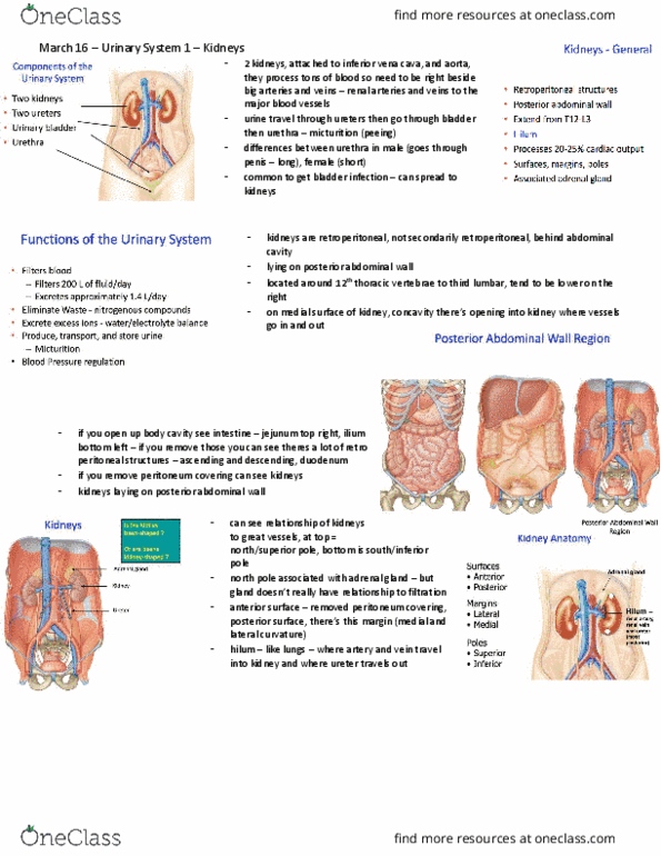
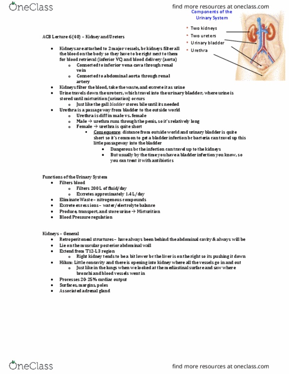
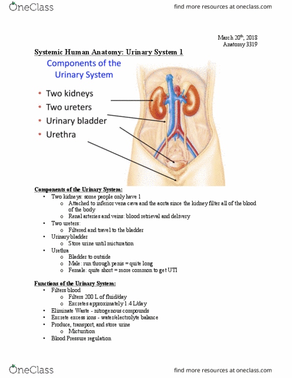
Describe the gross anatomy of the structures imperative for, and their underlying role in the micturition reflex. Justify the location where stones may lodge in the urinary tract. Bladder & urethra: describe the gross anatomy of the bladder, compare and contrast the anatomy of the male and female urethrae, explain the normal neuronal control of the bladder and its coordination during micturition. Filters blood: almost 200l/day, excretes approx. Tx: typically left in body, transplant goes in pelvis connected to common iliac vessels. Innermost layer: attached to surface of kidney, protects kidney, perirenal fat, protective mass of fatty tissue, cushioning function, renal fascia, dense, irregular ct, anchors kidney & adrenals in abdomen, pararenal fat, protective mass of fatty tissue, cushioning function. Duct draining the renal pelvis into the bladder. Enters bladder at a sharp angle acting as a natural valve. Note 3 points of constriction kidney stones get lodged. Intravenous pyelogram showing a kidney stone lodged in the ureter (arrow)