Anatomy and Cell Biology 3319 Lecture Notes - Lecture 39: Pleural Cavity, Muscles Of Respiration, Pneumothorax

6 May 2019
School
Department
Professor
Document Summary
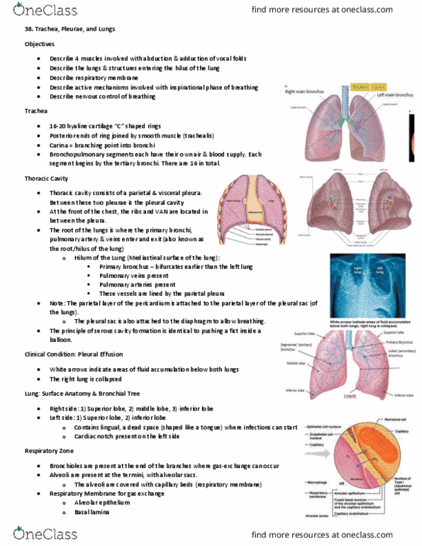
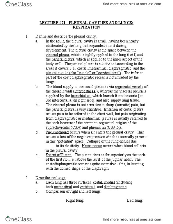
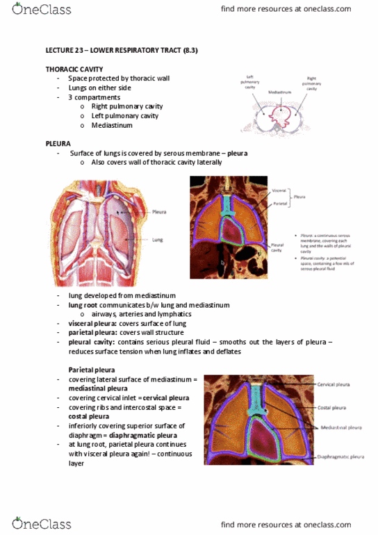
Respiratory system ii the lower resp tract & breathing. = always lower than atm. pressure = keeps lungs filled w/in p. c. , despite the tendency of lungs to recoil due to their inherent elasticity. 2: lungs, lungs occupy the bilateral portions of the thoracic cavity, separated by mediastinum. Lungs in the anatomy lab: spongy, styrofoam-like feel. Lungs cont"d: pyramid-shaped, apex, base, surfaces (3), costal, diaphragmatic, mediastinal, borders (3), anterior, posterior. Lobes of the lungs: lobes, right (larger) = 3 lobes superior, middle, inferior, left (smaller) = 2 lobes superior, inferior, separated by fissures, oblique, horizontal. The hilum (aka root of the lung: hilum, location: on mediastinal surface of each lung, where parietal & visceral pleura meet, where all structures enter/exit lung, structures at hilum, pulmonary arteries, pulmonary veins, air passages (bronchi) What are the clinical implications of such a large capillary bed: answer: insoluble material (ex. cell clumps, clots) present in blood can become lodged in pulmonary capillaries.