Anatomy and Cell Biology 4451F/G Lecture Notes - Lecture 5: Olfactory Bulb, Olfactory Bulb Mitral Cell, Extracellular Fluid

12 Sep 2018
School
Department
Professor
Document Summary
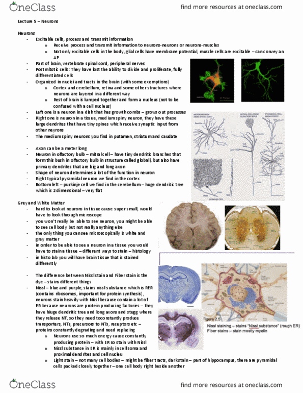
Neurons come in all different shapes, typically, the motor neurons have big soma and lots of dendrites with a long axon. Mitral cells have really tiny dendritic branches that form a bush within olfactory bulb and a structure called globuli. Shape of neurons usually defines the function of the neuron. In order to be able to see a neuron in a tissue, you have to stain the tissue. There are different types of stains, fiber stains and nissl stain: nissl stai(cid:374)i(cid:374)g stai(cid:374)s (cid:862)nissl su(cid:271)sta(cid:374)(cid:272)es(cid:863) (rough er) nissl stain mostly cell somas, nucleus, and maybe dendrites, fiber staining stains mostly myelin. Because they contain a lot of endoplasmic reticulum. White dots are the heavily stained myelinated axons. What you see under microscope depends on what you have stained for. When looking at structures, you can use hippocampus as the landmark. These dark lines in the hippocampus are called dentate gyrus (dg).