Anatomy and Cell Biology 3309 Lecture Notes - Lecture 27: Pars Tuberalis, Pituitary Gland, Supraoptic Nucleus

22 May 2018
School
Department
Professor

Histology 3309
Pituitary Gland
Learning Objectives
1. name the two main divisions of the pituitary gland and list their parts
2. describe the development of the pituitary, including the derivation of its divisions.
3. describe the blood supply of the pituitary gland
4. name the five main cell types in the pars distalis and list the hormones they secrete
5. explain the functions of anterior pituitary hormones
6. name the components of the neurohypophysis
7. name the hormones released in the pars nervosa and explain their
function.
8. describe the significance of pituicytes and Hering bodies
9. explain the relationship between hypothalamic nuclei and pituitary
hormone release.
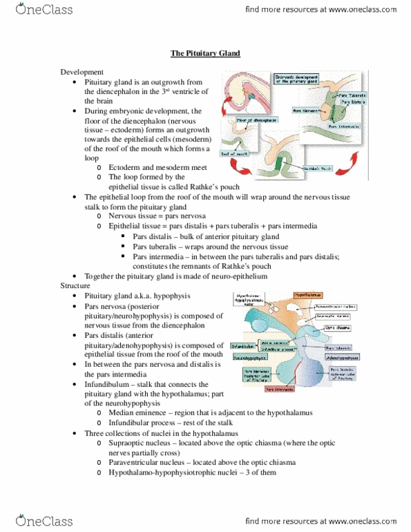
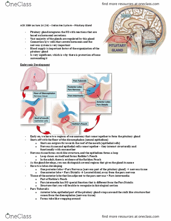
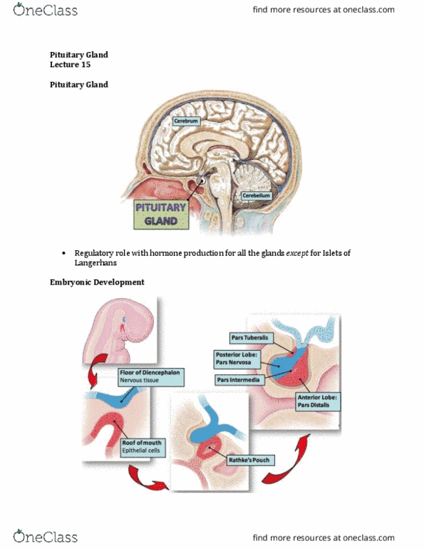
Embyronic Development
- there are 2 regions of our anatomy that come together to form our
pituitary gland:
o floor of diencephalon (neural epithelium) forms an outgrowth towards the roof of the
mouth
o roof of the mouth is endoderm (future mouth region) – epithelial cells
- so epithelial cells and nervous tissue come together and interact structurally and functionally
with one another
- so the nervous tissue (blue) forms stock like structure whereas the epithelium forms the loop - a
loop that actually closes on itself and forms rathke’s pouch
- later on in the adult, you will see evidence of rathke’s pouch
- as the gland develops, we can distinguish several regions:
- we have 2 lobes developing:
o posterior lobe (aka pars nervosa)
o anterior lobe (aka pars distalis)
▪ there is also a pars intermedia →part of rathke’s pouch that lies adjacent to the
pars nervosa
▪ has no special function that is different from the pars distalis
- pars tuberalis: red tissue
o the epithelial part of the pituitary gland wraps around the stock like structure that
comes from the diencephalon (nervous tissue)
- this is a pic of the mature/developed pituitary gland
- since the pituitary gland is in part made of nervous tissue, it lies direcetly under part of the brain
that later develops into the hypothalamus
find more resources at oneclass.com
find more resources at oneclass.com

- so the hypothalamus and the pituitary gland communicate bc part of the pituitary gland is
nervous tissue e
- in the hypothalamus, there are 3 regions called nucleus (regions where there are neuron cell
bodies)
o these concentrations of cell bodies develop the capacity to produce hormone
o so rather than signaling electrically, they produce signaling molecules that are not
designed to stimulate other nerves, but to be released into the bloodstream
- these nuclei are called:
o paraventricular nucleus
o supraoptic nucleus – nerve cells that are sitting over a region where the optic nerve
crosses (optic chiasma)
o hypothalamo-hypophsiotropic nuclei
- these 2 regions of nuclei send axons right into the pars nervosa
- the pars nervosa (aka posterior lobe of pituitary) is aka neurohypophysis
- so another name for the pituitary gland is hypophysis
- these axons pass through this stock that is also called infundibulum (this is divided into 2
regions):
o infundibular process
o median eminence
▪ adjacent to the brain
▪ functionally, this is the location where axons (nerve cell processes) end and
these nerve cell processes come from the 3rd nucleus
▪ this tells you that the nuclei are located in the hypothalamus and they feed the
hypophysis – they feed the anterior lobe of the hypophysis
- so the anterior lobe of the hypophysis – pars distalis (made of epithelial tissue) is also called
adenohypophysis
- pars tuberalis: extension of the adenohypophysis which wraps around on either side of the
infundibulum
- know these terms and understand how the nervous tissue relates to the epithelial tissue!!
- blood comes in to the pituitary gland from 2 locations:
o inferior hypophysial artery that comes from below
▪ that feeds into a capillary network within the pars nervosa
o superior hypophysial artery
▪ supplies the pars distalis
find more resources at oneclass.com
find more resources at oneclass.com

▪ this artery does not directly go into the pars distalis, it first forms a capillary
network in the median eminence of the infundibulum
▪ this is a primary capillary plexus
▪ then the blood flows through veins and venules into the pars distalis where it
forms a secondary capillary network
▪ this is another example of a portal system (pars distalis is supplied by a
secondary capillary network) →this is called the hypothalamohypophysial
portal system
- paraventricular nucleus and supraoptic nucleus send axons into the pars nervosa and release
hormones into the capillary network that is made by the inferior hypophysial artery
- these hormones are oxytocin and ADH
o these hormones are produced in the hypothalamus (where the nerve cell bodies are)
and then they are sent down the axons into the pars nervosa and dumped into the
capillary network
- for the hypothalamo-hypophsiotropic nuclei, they end in the median eminence – so these
processes end there and deliver into the primary capillary network of the superior hypophysial
artery, releasing inhibitory hormones that travel through the portal system into the anterior
lobe of the pituitary gland (yellow squares)
o these are the factors that stimulate the epithelial cells that live in the anterior pituitary
gland to produce a variety of hormones and factors
- hormones produced by the epithelial cells are shown in the green box
find more resources at oneclass.com
find more resources at oneclass.com
Document Summary
Note: the hormones that are released in the pars nervosa are produced in the hypothalamus by those neurons but the hormones that are produced in the ant lobe are both produced and released in that same location. We have corticotrophs that produce adrenocorticotrophic hormone (acth) Lactotrophs (aka mammotrophs) produce prolaction (important for milk production in the mammary gland) Diff cell types live next to eachother. Acidophilic mammotroph (aka lactotroph) and the basophilic corticotroph that can be distinguished by the size of their secretory granules. You don"t really need to know t(ese 2 sl)des. Cells of the adenohypophysis and the hormones they produce. Chromophobes are chromophils that have secreted stuff (cid:523)they don"t have lots of content so they don"t stain(cid:524) Then we have chromophils which are divided into 2 cells: basophils, acidophils the activity of the cells in the anterior pituitary gland (ex: basophilic gonadotroph) is regulated by the hypothalamus.