Anatomy and Cell Biology 3309 Lecture Notes - Lecture 7: Dense Irregular Connective Tissue, Hyaline Cartilage, Chondroitin Sulfate

3 Dec 2014
School
Department
Professor


Document Summary
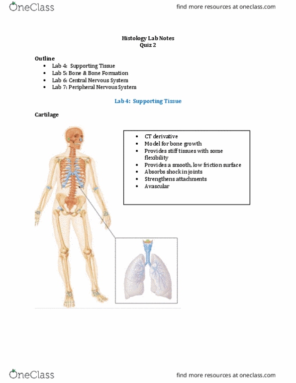
Cartilage is found throughout the body, and its function is based on its structure. Cartilage does not heal well because it is avascular. Three kinds of cartilage: hyaline cartilage: glassy smooth appearance in microscope; found in majority of joints, elastic cartilage: hyaline cartilage with elastic fibers and plates for flexibility and memory, fibrocartilage: hyaline cartilage embedded in d. r. c. t. 3-5% is cells, rest is ecm (water, collagen, proteoglycans, glycoproteins) General organization of cartilage tissue, specifically hyaline cartilage: avascular, chondrocytes surrounded by matrix, matrix around chondrocytes = territorial matrix (stains deeper blue because more sulfated. Gags in this area, more basophilic); just made by chondrocytes: matrix further out from chondrocytes = interterritorial matrix (stains lighter blue because less sulfated groups, surface covering of dense irregular connective tissue called perichondrium. In the chondrogenic layer, chondrocytes will undergo appositional growth (surface growth: note: bone is calcified and so cannot undergo interstitial growth major. Perichondrium layer they would not rub together as well.