Anatomy and Cell Biology 3319 Lecture Notes - Lecture 38: Gastrointestinal Tract, Tunica Externa, Lamina Propria

1 May 2018
School
Department
Professor

Lecture 038: Swallowing, Stomach and Formation of Mesenteries
Objectives
● Describe the structures/actions involved with swallowing.
● Describe the arrangement of the tunica adventitia, muscularis, submucosa and mucosa
in the esophagus.
● Describe the different regions and cell types of the stomach.
● Describe the arterial blood supply to the stomach via branches of the celiac trunk.
● Describe the formation and functions of the mesenteries.
○ Give 5 examples of mesenteries.
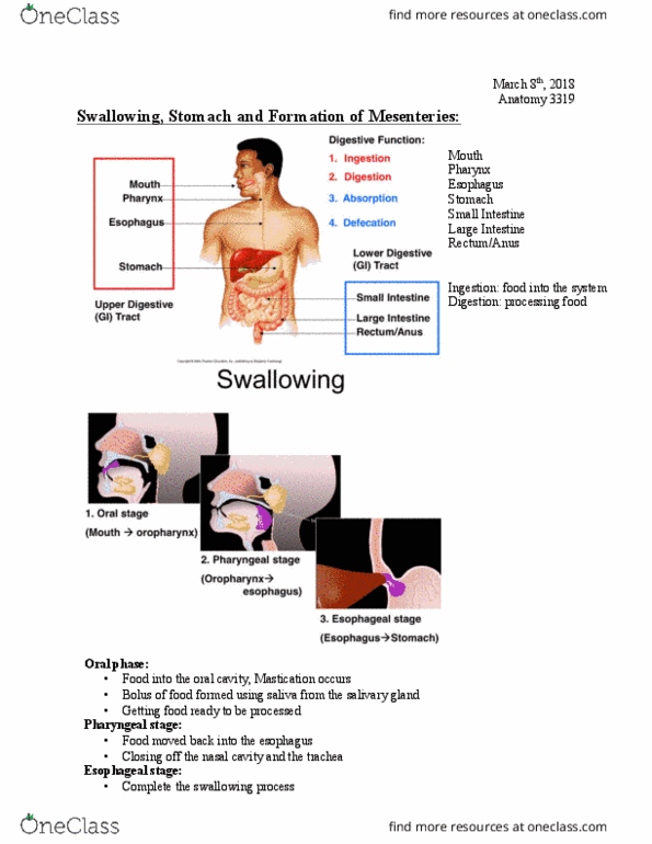
Ingestion: taking food into the system
Digestion: processing food so nutrients can be extracted
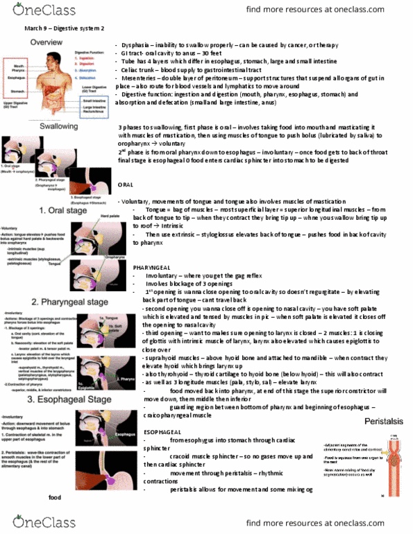
Swallowing Food
1. Oral stage
● Food is taken into the oral cavity
● Through the process of mastication and saliva a bolus of food is formed
● Mouth -> oropharynx
● Voluntary
● Action:
○ Pushes food together into a bolus shape
○ Tongue elevates and pushes food against the hard palate and backwards into
the oropharynx
● Muscles involved:
○Intrinsic muscles of the tongue
■ Superior longitudinal muscles
● Curl the tongue and pushes the food back
○Extrinsic muscles of the tongue
■ Originates outside of the tongue and
■Styloglossus and palatoglossus
■ Pushes food back until it reaches the pharynx
2. Pharyngeal stage
● Food is moved from the oropharynx into the esophagus
● Must close off the nasal cavity and the trachea when you swallow food
● Oropharynx -> esophagus
● Involuntary
○ Most actions are part of the gag reflex
● Action:
○Blockage of 3 openings:
■Blockage of the oral cavity
● Continuous elevation of the tongue in the oral cavity
find more resources at oneclass.com
find more resources at oneclass.com

● Tongue is pushed up against the hard palate so that food has
nowhere to go but down the throat
■Blockage of the nasal cavity
● Elevation of the soft palate
● Occurs through the actions of the levator palatini and tensor
palatini
■Blockage of the trachea
● Elevation of the larynx
○ This cause the epiglottis to fold over the laryngeal inlet
(covers the opening of the trachea)
● Occurs through the actions of
○ Muscles above the hyoid bone
■ Suprahyoid muscle
■ Thyrohyoid muscle
○ Vertical muscles of the laryngopharynx
■ Palatopharyngeus,
■ Stylopharyngeus
■ Salpingopharyngeus
○Contraction of the pharynx forces the bolus into the esophagus
■ Occurs by the sequential actions of the superior, middle, and inferior
constrictors
3. Esophageal stage
● Involuntary
○ Causes downward movement of bolus through esophagus and into stomach
● Actions:
○ Contraction of skeletal muscle in the upper part of esophagus
■ Contract to force food down the esophagus
○Peristalsis
■ Sequential wave-like contraction of skeletal muscle and smooth muscles
in the lower part of the esophagus (and the rest of the alimentary canal)
● Moves the food down the esophagus
■ Adjacent segments of the alimentary canal relax and contract
● Food is squeeze from one organ to the next
● Note: some mixing of food (by segmentation) occurs as well
● Food moves to the stomach pass the cardiac sphincter
● Completes the swallowing process
find more resources at oneclass.com
find more resources at oneclass.com

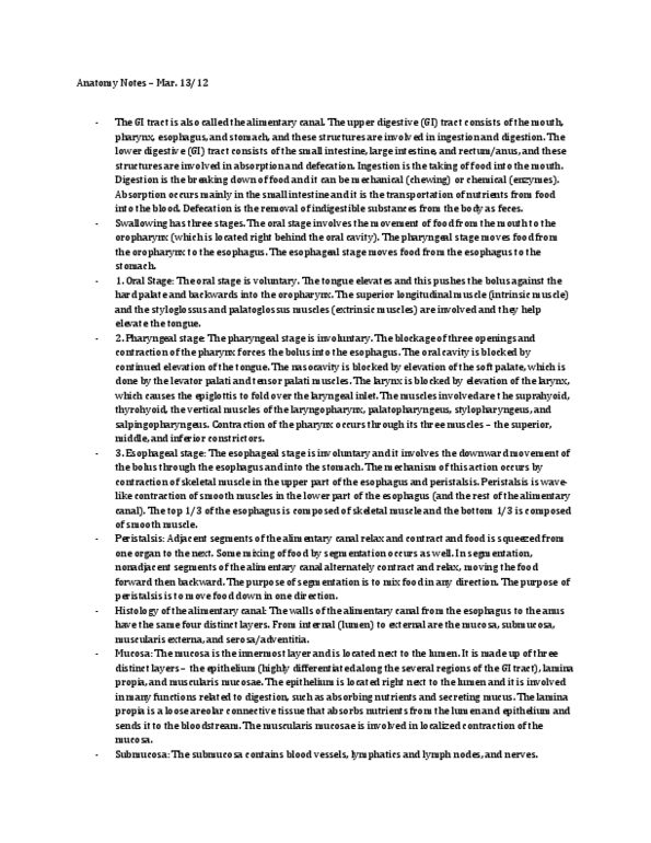
Histology of the Alimentary Canal
● The walls of the GI tract (from esophagus to the anus) have the same four distinct layers
○Mucosa
■ Innermost layer
■ Composed of:
● A layer of epithelium
○ Highly differentiated along the several region of the GI tract
● Lamina propria
○ Basement membrane
● Muscular mucosas
○ Thin muscular layer
○ Submucosa
■ Lots of connective tissue
■ Contains blood vessels
● Nourishes the esophagus
■ Lymphatics and lymph nodes
■ Nerves
● Controls the parastais
○ Muscularis Externa
■ Composed of a layer of circular muscles (inner) and a layer of longitudinal
muscle (outer)
○ Serosa/Adventitia
■ Outermost layer
■ Attaches the GI tract to the surrounding structures
Histology of the esophagus
Layer Esophagus
Mucosa Stratified squamous epithelium
Submucosa Areolar connective tissue
Muscularis externa 2 layers:
● Inner circular
● Outer longitudinal
○ Superior ⅓:skeletal
○ Middle ⅓: skeletal + smooth
○ Inferior⅓: smooth
Adventitia/Serosa Adventitia
● Connective tissue layer that anchors
organ in place
Esophagus
find more resources at oneclass.com
find more resources at oneclass.com
Document Summary
Lecture 038: swallowing, stomach and formation of mesenteries. Describe the arrangement of the tunica adventitia, muscularis, submucosa and mucosa in the esophagus. Describe the different regions and cell types of the stomach. Describe the arterial blood supply to the stomach via branches of the celiac trunk. Describe the formation and functions of the mesenteries. Digestion: processing food so nutrients can be extracted. Food is taken into the oral cavity. Through the process of mastication and saliva a bolus of food is formed. Pushes food together into a bolus shape. Tongue elevates and pushes food against the hard palate and backwards into the oropharynx. Curl the tongue and pushes the food back. Pushes food back until it reaches the pharynx: pharyngeal stage. Food is moved from the oropharynx into the esophagus. Must close off the nasal cavity and the trachea when you swallow food. Most actions are part of the gag reflex. Continuous elevation of the tongue in the oral cavity.