Nursing 3920A/B Lecture Notes - Lecture 3: Antiplatelet Drug, Systolic Geometry, Ductus Arteriosus

24 Apr 2018
School
Department
Course
Professor
Document Summary
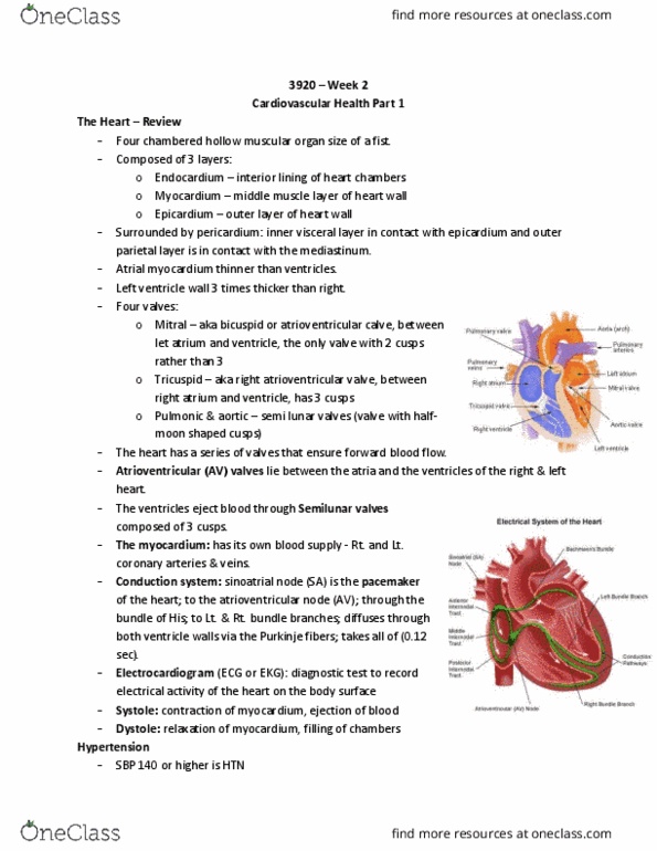
Learn about the prevention, management and experience of heart disease. Cad: anatomy review, htn, angina, myocardial infarction, assessment & management. Four chambered hollow muscular organ size of a fist. Composed of 3 layers: endocardium, myocardium, and epicardium. Surrounded by pericardium: inner visceral layer in contact with epicardium and outer parietal layer is in contact with the mediastinum. Epicardium = outer layer of the heart wall: atrial myocardium thinner than ventricles. Left ventricle wall 3 times thicker than right. Four valves: mitral & tricuspid; pulmonic & aortic. Tricuspid valve = also called (right atrioventricular valve) b/t right atrium & ventricle, has 3 cusps. Pulmonic & aortic valves = are semilunar valves. The heart has a series of valves that ensure forward blood flow. ventricles of the right & left heart. The ventricles eject blood through semilunar valves composed of 3 cusps. The myocardium: has its own blood supply - rt. and lt. coronary arteries & veins.