BIOL 2021 Lecture Notes - Dark Field Microscopy, Optical Microscope, Bright-Field Microscopy


Document Summary
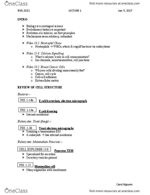
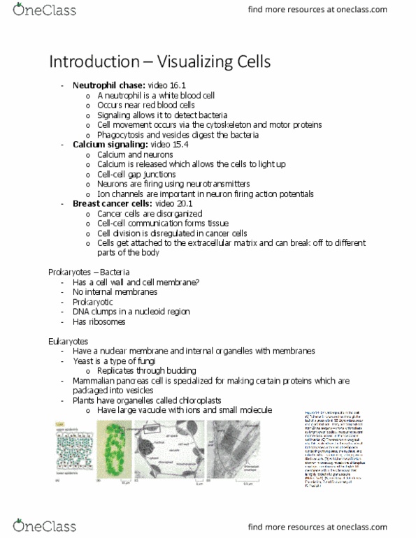
Review cell structure (chapter 8 & 9) No internal membrane- prokaryotes only have plasma membrane. Bacteria have cell wall, cell membrane, ribosomes and enzymes inside. Cell walls- bacteria, plants, fungi are all different. Bacteria and mitochondria are usually smallest objects whose shape we can discern in the light microscope. Bright field (full light spectrum): normal light microscope in which the image is obtained by simple transmission of light through the object being viewed: phase contrast. No need for stain; transparent cells become visible. 4 types of light microscopy-(a) bright field microscopy, (b)phase-contrast, (c) narmarski differential-interference-contrast microscopy, (d) dark field microscopy: stain cell. Involves killing cells, slicing thin cells and staining them. Sliced cells are treated with a fixative and then embedded in a supporting medium before sectionin. Cells are usually invisible under ordinary light microscope, so they often must be stained with organic dye that have a specific affinity for particular subcellular componenets- different stains for different components.