KINE 3012 Lecture Notes - Lecture 21: Endoplasmic Reticulum, Ryanodine, Electrocardiography

12 Feb 2018
School
Department
Course
Professor
Document Summary
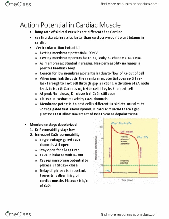
After contraction, ca2+ is pumped back into sr: time of contraction & ap are same, involves l-type calcium channels. Its allows prolonging of refractory period and links excitation & contraction. Calcium also binds to sr channels (ryanodine receptor)- it"s a ligand gated channel. It allows release of calcium from sarcoplasmic reticulum. Amount of calcium released from sr is much more. Calcium can now allow acting & myosin contraction just like in skeletal muscle: to increase strength of cardiac muscle, more ca2+ is released as it allows more actin-myosin contraction. In skeletal muscle, strength is based on activation of muscle fibers & motor units. Excitation- contraction coupling: l-type calcium channels in t-tubules which allows small amount of calcium to enter. This increases cytosol calcium through increase by sr. This allows actin-myosin interaction to form cross bridge to cause contraction. The electrocardiogram (ecg: how we measure electric activity, sum of all ap, p-wave: atrial depolarization.