PSYC 2220 Lecture Notes - Lecture 4: Functional Magnetic Resonance Imaging, Positron Emission Tomography, Hemoglobin

18 Jan 2016
School
Department
Course
Professor
Document Summary
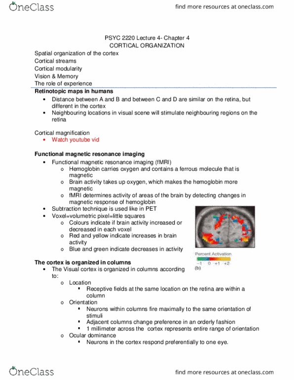
Retinotopic maps in humans: distance between a & b and between c & d are similar on the retina. Brain imaging techniques: cortical mapping in humans has been revealed using imaging techniques. Pet fmri: pet (positron emission tomography) Person is injected with a harmless radioactive tracer. Changes in blood flow show changes in brain activity: subtraction method, brain activity is determined by . Subtracting the control activity from the stimulation. Activity fmri (functional magnetic resonance imaging: hemoglobin carries oxygen & contains a ferrous molecule that is. Which make the hemoglobin more magnetic: fmri determines activity of areas of the brain by detecting changes in. Subtraction technique is used like in pet the cortex is organized in columns: the visual cortex is organized in columns according to . Receptive fields at the same location on the retina are. Neurons within columns fire maximally to the same orientation of stimuli. Adjacent columns change preference in an orderly.