PHSL232 Lecture Notes - Lecture 5: Purkinje Fibers, Atrioventricular Node, Depolarization

Document Summary
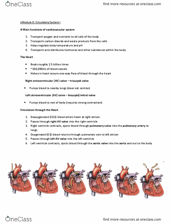
Explain the generation and conduction of the ap in the heart. Explain how the action potentials are generated in the nodal cells and myocardial cells. State the difference between nodal and ventricular action potentials. Understand the basic concepts of the ecg. Explain some of the important clinical issues associated that can be diagnosed using an ecg. Nodal tissue: (sa node and av node- structurally very similar) Contains small round cells with little or no contractile proteins. Specialised for the generation and conduction of action potentials in the atria. Action potentials spontaneously generated in the sa node. Ap can travel from cell to cell via gap junctions- organised along preferential pathways, determined by location of intercalated discs. Main conduction pathways comprise groups of specialised cells. To get to the ventricles, ap has to pass through av ring ( via av node) Sa node generates ap at about 100-110/min (hr under parasympathetic control- regulates normal heart rate)