BIOL 030 Lecture Notes - Lecture 23: Fovea Centralis, Aqueous Humour, Optic Disc

17 Sep 2020
School
Department
Course
Professor
Document Summary
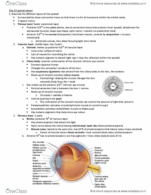
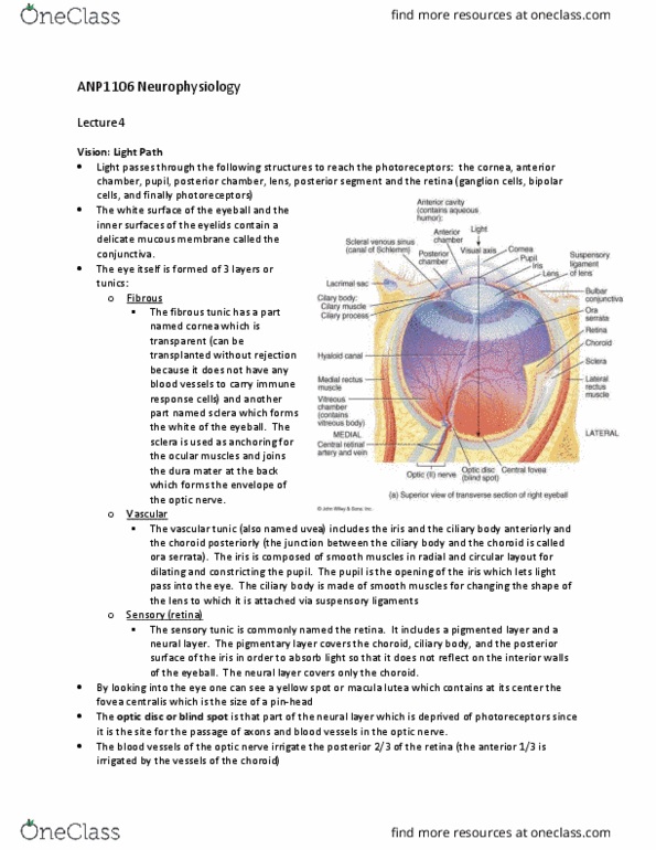
Three layers (tunics) that form the wall of the eyeball. Middle vascular layer = tunica vasculosa (uvea) Cornea: transparent region of modified sclera in front of eye that admits light. Choroid: highly vascular, deeply pigmented layer behind retina. Ciliary body: extension of choroid; a muscular ring around lens. Supports lens and iris: secretes aqueous humor. Iris: colored diaphragm controlling size of pupil (opening) Transparent elements that admit light, refract light rays, and focus images on retina. Opthalmoscope tool used to examine retina and blood vessels. Macula lutea: patch of retina on visual axis of eye (3 mm diameter) Fovea centralis: center of macula, finely detailed images due to packed receptor cells. Optic disc: no receptor cells blind spot. The body has 4 principal mechanisms of communication between cells: electrolytes to move from cell to cell. Gap junctions: pores in cell membrane allow signaling molecules, nutrients, and. Neurotransmitters: released from neurons to travel across synaptic cleft to second.