PSYC 2012 Lecture Notes - Lecture 21: Frontal Eye Fields, Bipolar Neuron, Amacrine Cell

Document Summary
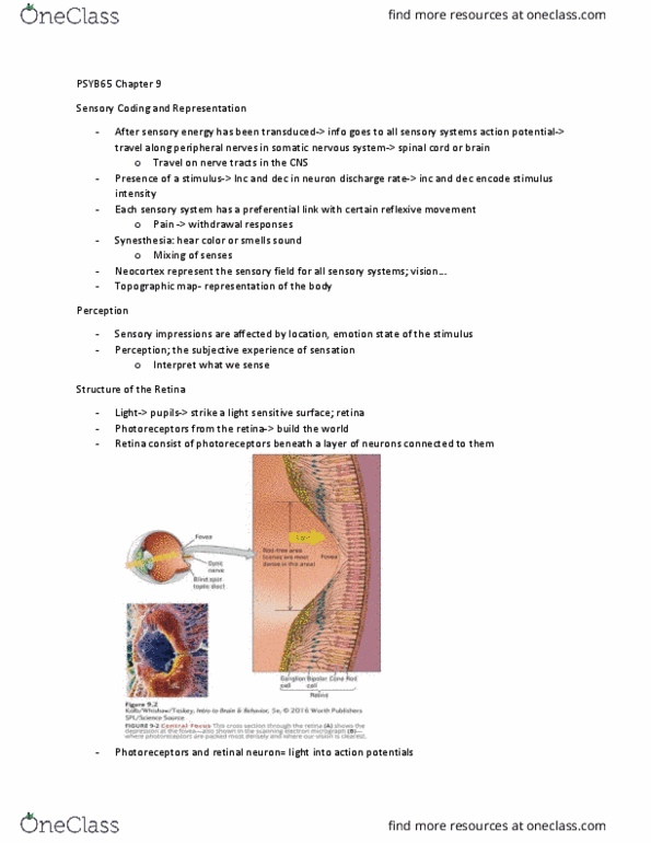
Consists of sensory receptors (photoreceptors) and the sensory neurons that synapse with them. Blind spot - region containing no photoreceptors because the sensory neuron axons exit the eye here as the opti(cid:272) (cid:374)er(cid:448)e (cid:271)ut the other eye, a(cid:374)d photore(cid:272)eptors arou(cid:374)d ea(cid:272)h (cid:271)li(cid:374)d spot, (cid:862)fill i(cid:374)(cid:863) missing visual info. Saccades are controlled by the frontal eye fields (cortex) and the superior colliculus (midbrain) They are responsible for color vision and our ability to see fine detail. Rods are more numerous than cones and are more sensitive to dim light. Only one type of light absorbing pigment. 3 different types of light absorbing pigment. Photoreceptors contain a special pigment molecule called rhodopsin. When light strikes rhodopsin, it activates a g-protein. The activated g protein closes a na+ channel. Closing the na+ channel means less (+) charge flowing into the cell, hyperpolarizing it. Hyperpolarization means decreased release of glutamate by the photoreceptor onto bipolar cells.