HSCI 301 Lecture Notes - Lecture 20: Resting Potential, Propranolol, Verapamil

1 Dec 2016
School
Department
Course
Professor
Document Summary
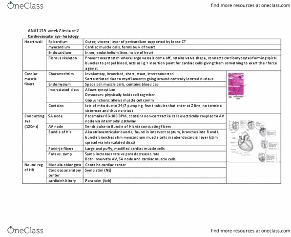
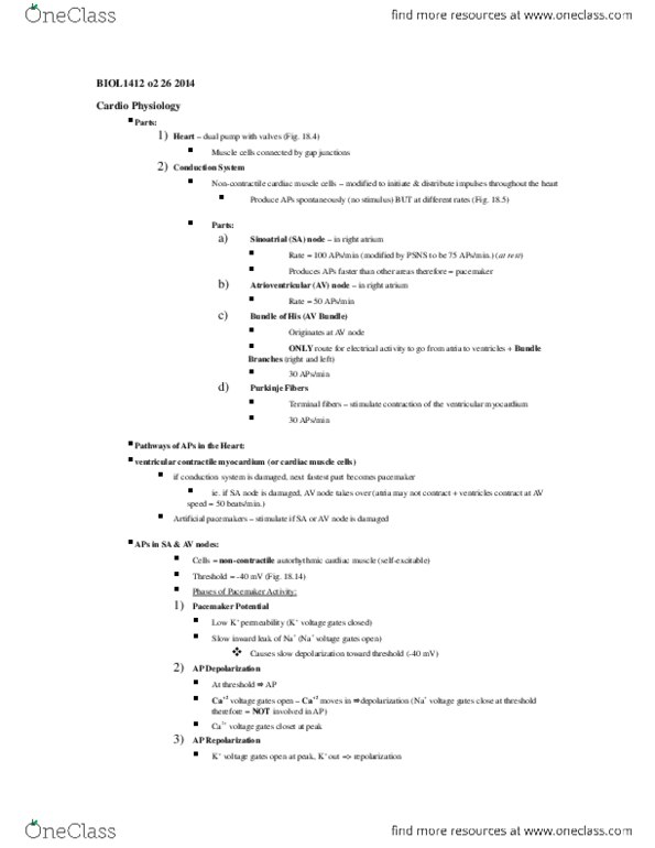
Chapter 20: antiarrythmics: understand the subtle differences between cardiomyocytes and cardiac. Pacemaker cells (modified cardiomyocytes), and how the heart electrically behaves as one large cell. Cardiomyocytes: heart cells of atria (pumped in) & ventricles (pumped out) Cardiac pacemaker cells (modified cardiomyocytes): carries the impulses through the heart: kinds: Sa node (primary pacemaker) makes average resting hr is 70 bpm. Receive & respond to impulses from cns (lets you adjust resting. Impulse goes from depolarization of sa node > atrium > av node > bundle of his. This is the pattern of an ecg: understand the ion movement associated with the different phases of the cardiac. Phase 0: depolarization (na+ comes into cell) Phase 1: overshoot (na+ comes into cell) Phase 2: plateau (ca2+ comes into cell) Phase 4: resting membrane potential again: associate cardiac cell phase inhibition to particular antiarrhythmic drug.