PSYC 372 Lecture Notes - Lecture 4: Positron Emission Tomography, Magnetic Resonance Imaging, Ct Scan

15 Feb 2018
School
Department
Course
Professor
Document Summary
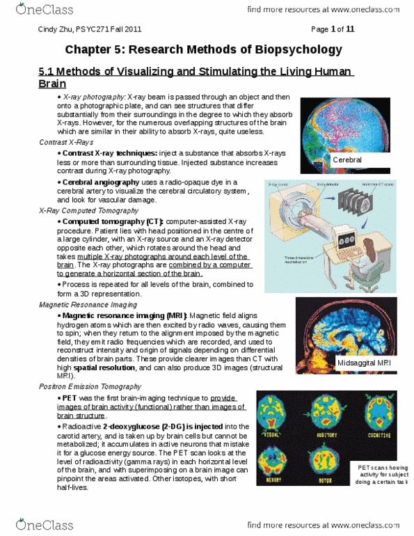
Lecture 4 notes: structural imaging, computerized tomography (ct) scanning. Brain tissue does not show up well on a traditional x-ray. Inject a substance (iodine) that absorbs x-rays to enhance contrast and allow visualization of brain tissue. Developed in the early 1970s: magnetic resonance imaging (mri) A general technique that can be used to determine the amount of certain types of atoms in different locations in the body. In biopsychology, used to measure to the hydrogen atoms within the water and fat of the brain. The protons of hydrogen atoms are activated by high-frequency magnetic waves. Alignment of the atoms generates a detailed structural image. Can be used to generate a series of 2d or 3d images. Static magnetic field: protons of the body will align with the static magnetic field. Radiofrequency pulse: repeatedly turned on and off, protons will change their state of excitability (on) and return to normal by releasing the energy (off)