PSYC 271 Chapter Notes - Chapter 5: Transcranial Magnetic Stimulation, Magnetic Resonance Imaging, Cerebral Angiography

12 Mar 2014
School
Department
Course
Professor
Document Summary
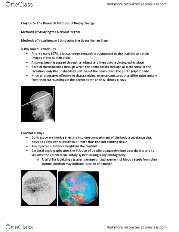
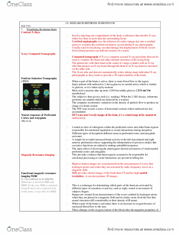
5. 1 methods of visualizing and stimulating the living human. Brain: x-ray photography: x-ray beam is passed through an object and then onto a photographic plate, and can see structures that differ substantially from their surroundings in the degree to which they absorb. However, for the numerous overlapping structures of the brain which are similar in their ability to absorb x-rays, quite useless. Contrast x-rays: contrast x-ray techniques: inject a substance that absorbs x-rays less or more than surrounding tissue. Injected substance increases contrast during x-ray photography: cerebral angiography uses a radio-opaque dye in a cerebral artery to visualize the cerebral circulatory system, and look for vascular damage. X-ray computed tomography: computed tomography (ct): computer-assisted x-ray procedure. Patient lies with head positioned in the centre of a large cylinder, with an x-ray source and an x-ray detector opposite each other, which rotates around the head and takes multiple x-ray photographs around each level of the brain.