ANTR 350 Lecture Notes - Lecture 4: Internal Sphincter Muscle Of Urethra, Urinary Meatus, Urethral Sphincters

Document Summary
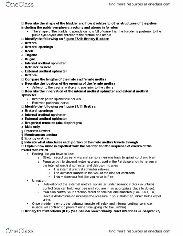
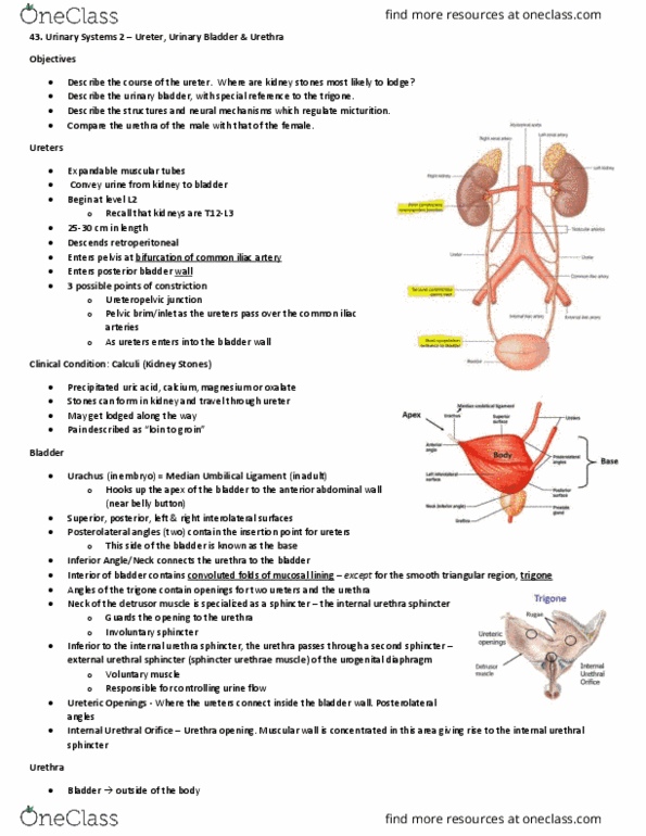
Remains retroperitoneal, blood will be supplied through branches from nearby arteries and drainage will occur in companion (same-named) veins. Referred pain from kidney and ureters to t10-t12 dermatomes. Shape: empty-upside down pyramidal full- distends superiorly until oval. Females- anterior to vagina and anteroinferior to uterus. Males- anterior to rectum, superior to prostate gland. Female: 3-5 cm, opens outside of body at external urethral orifice in female perineum. More often in females than males because shorter urethra and urethral orifice is closed to anus and vaginal opening. Starts with inflammation of urethra and infection spreads to bladder and then to kidneys through ureters. Innervation of detrusor, internal urethral sphincter, external urethral sphincter. Detrusor and internal urethral sphincter- smooth muscles controlled by ans. External urethral sphincter- skeletal muscle under voluntary control. Parasymp- stimulates micturition by relaxing internal urethral sphincter and contracting detrusor(parasymp makes you pee) Symp- inhibits micturition by contracting internal sphincter and inhibiting contraction of detrusor.