BIOL 129 Lecture Notes - Lecture 12: Cricoid Cartilage, Bronchopulmonary Segment, Hyoid Bone

Document Summary
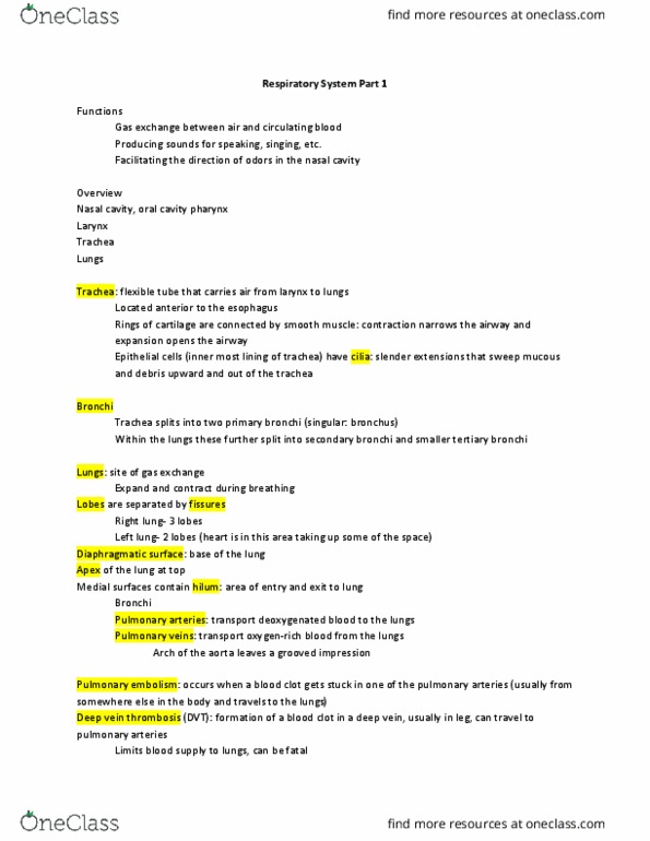
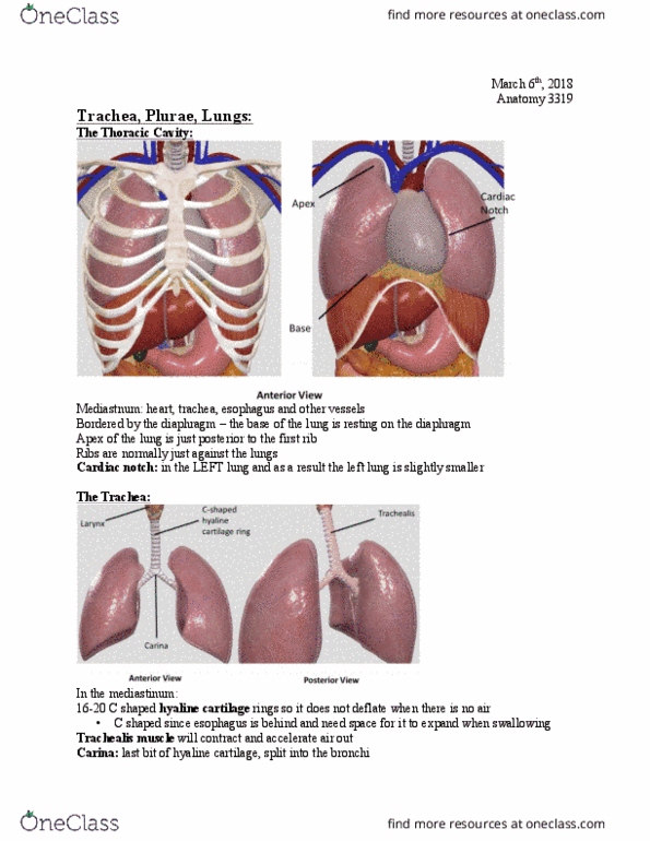
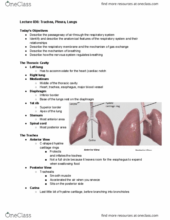
Gas exchange between air and circulating blood. Facilitating the detection of odors in the nasal cavity. Flexible tube, carries air from larynx to lungs. Located anterior to the esophagus, posterior to the heart. Rings of cartilage provide structure, connected by smooth muscle. C shaped means they don"t close in the back. Cilia line mucosa of trachea cilia are covered with mucus. Treach is lined with mucus producing epithelium and ciliated cells. Cilia created mucociliary escalator - to pharynx. Trachealis muscle spans in c-shaped cartilage ring. Trachea splits into two primary bronchi (singular = bronchus) Within the lungs, these further split into secondary bronchi, and smaller tertiary bronchi. Heart has to be fitted into the left side. Medial surfaces contain the hilum : area of entry and exit to lungs. Each tertiary bronchus supplies air to lung segment called a bronchopulmonary. Bronchioles are small branches or tertiary bronchi and feed into alveolar ducts. Small alveolar ducts branch off of bronchioles.