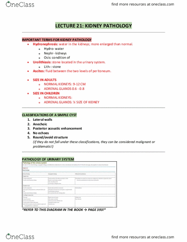
BIOL 171 Lecture Notes - Lecture 22: Calcinosis, Hydronephrosis, Connective Tissue

Document Summary
Get access


Related Documents
Related Questions
Case-3: Patient History
P.W. is 23 years old. He was the victim of a hit-and-run auto-pedestrian accident and suffered multiple abrasions, a concussion, and a deep laceration of his left thigh. He was discovered approximately 2 hours after the incident and is now in the emergency department. P.W.âs vital signs and hematocrit suggest that he has had a blood loss of about 2500 ml. A urinary catheter is inserted to monitor urine output, and fluid resuscitation is initiated while his wounds are cleaned and sutured. The urine output is averaging 15 ml/hr, with a high urine osmolality and low urine sodium.
What type of renal failure is P.W. likely developing? (select all that apply)
| Intrarenal acute renal failure |
| Prerenal acute renal failure |
| Postrenal acute renal failure |
| Chronic renal failure Based on your answer for the previous question, what is the best therapy for preventing this from occurring? (select all that apply)
In addition to urine output, what laboratory data should be monitored to assess changes in P.W.âs renal function? (select all that apply)
|
P.W. is 23 years old. He was the victim of a hit-and-run auto-pedestrian accident and suffered multiple abrasions, a concussion, and a deep laceration of his left thigh. He was discovered approximately 2 hours after the incident and is now in the emergency department. P.W.âs vital signs and hematocrit suggest that he has had a blood loss of about 2500 ml. A urinary catheter is inserted to monitor urine output, and fluid resuscitation is initiated while his wounds are cleaned and sutured. The urine output is averaging 15 ml/hr, with a high urine osmolality and low urine sodium.
Analyze this case study and answer the next four questions that follow.
Case-3: Question-4
If P.W.âs renal function does not return to normal, but continues to be diminished, what renal disorder might develop? (select all that apply)
| A) He may develop nephrotic syndrome |
| B) | He may develop renal calculi. |
| C) | He may progress to acute tubular necrosis. |
| D) | He may develop pyelonephritis |
