KAAP309 Lecture Notes - Lecture 15: Nasolacrimal Duct, Lacrimal Apparatus, Medial Rectus Muscle

18 May 2018
School
Department
Course
Professor

Ch. 15A Vision
I. The Eye and Vision
A. 70% of body's sensory receptors in eye
B. Visual processing by ~ half cerebral cortex
C. Most of eye protected by cushion of fat and bony orbit
II. Accessory Structures of the Eye
A. Protect the eye and aid eye function
1. Eyebrows
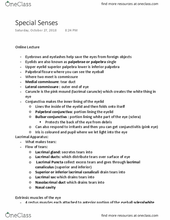
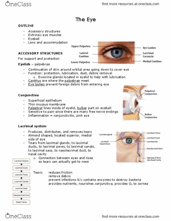
2. Eyelids (palpebrae)
3. Conjunctiva
a) Transparent mucous membrane
b) Produces a lubricating mucous secretion
c) Lines eyelids & covers sclera
4. Lacrimal apparatus
a) Makes & drains tears
b) Lacrimal gland
i. Above lateral end of eye
ii. Secretes tears
c) Nasolacrimal duct
i. Drains tears into nasal cavity
5. Extrinsic eye muscles
a) Six straplike extrinsic eye muscles
i. Originate from bony orbit; insert on eyeball
ii. Steer the eyes
b) Four rectus muscles
i. Superior, inferior, lateral, medial rectus
ii. Steer eye up, down, side-to-side
c) Two oblique muscles
i. Superior and inferior oblique
ii. Rotate eyeball about the central visual axis
III. Structure of the Eyeball
A. Wall of eyeball contains three layers
1. Fibrous
a) Outermost layer; dense avascular connective tissue
b) Two regions: sclera and cornea
i. Sclera
→ Opaque, white
→ Protects eyeball; anchors extrinsic eye muscles
→ Continuous with dura mater of brain posteriorly
ii. Cornea
→ Transparent anterior part of fibrous layer
→ Bends light as it enters eye
find more resources at oneclass.com
find more resources at oneclass.com

→ Numerous pain receptors contribute to blinking
and tearing reflexes
2. Vascular (uvea)
a) Middle (pigmented) layer
b) Three regions: choroid, ciliary body, and iris
i. Choroid region
→ Most of uvea; posterior portion of uvea
→ Supplies blood to all layers of eyeball
→ Brown pigment absorbs light to prevent light
scattering, which would cause unclear images
ii. Ciliary body
→ Ring of tissue surrounding lens: ciliary muscles
(parasympathetic) control lens shape, ciliary
zonule (suspensory ligament) holds lens in
position
iii. Iris
→ Colored part of eye
→ Pupil—central opening regulates amount of light
entering
• Sphincter pupillae (parasympathetic)
constrict
• Dilator pupillae (sympathetic) dilate
3. Inner (retina)
a) Originates as outpocketing of brain; 2 layers
i. Outer Pigmented layer
→ Single-cell-thick lining
→ Absorbs light and prevents its scattering
ii. Inner Neural layer
→ Transparent
→ Composed of three main types of neurons
• Photoreceptors, bipolar cells, ganglion cells
→ Signals spread from photoreceptors to bipolar
cells to ganglion cells
b) Quarter-billion photoreceptors: rods & cones
c) Optic disc (blind spot)
d) No photoreceptors where optic nerve leaves eye
B. Internal cavity filled with fluids called humors
C. Lens separates internal cavity into anterior and posterior segments (cavities)
IV. Photoreceptors
A. Rods
1. Dim light, peripheral vision receptors
2. More numerous, more light-sensitive than cones
3. No color vision or sharp images; numbers greatest at periphery
B. Cones
find more resources at oneclass.com
find more resources at oneclass.com
Document Summary
15a vision: 70% of body"s sensory receptors in eye, visual processing by ~ half cerebral cortex, most of eye protected by cushion of fat and bony orbit. Accessory structures of the eye: protect the eye and aid eye function, eyebrows, eyelids (palpebrae, conjunctiva, transparent mucous membrane, produces a lubricating mucous secretion, lines eyelids & covers sclera, lacrimal apparatus, makes & drains tears, lacrimal gland. Secretes tears: nasolacrimal duct, drains tears into nasal cavity, extrinsic eye muscles, six straplike extrinsic eye muscles, originate from bony orbit; insert on eyeball. Steer eye up, down, side-to-side: two oblique muscles. Structure of the eyeball: wall of eyeball contains three layers, fibrous, outermost layer; dense avascular connective tissue, two regions: sclera and cornea. Continuous with dura mater of brain posteriorly. Numerous pain receptors contribute to blinking and tearing reflexes: vascular (uvea, middle (pigmented) layer, three regions: choroid, ciliary body, and iris. Most of uvea; posterior portion of uvea. Supplies blood to all layers of eyeball.