MCB 150 Lecture Notes - Lecture 22: Fluorescence Microscope, Hybridization Probe, Nuclear Membrane

25 Dec 2015
School
Department
Course
Professor
Document Summary
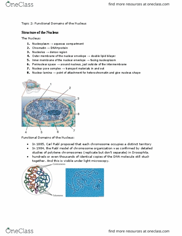
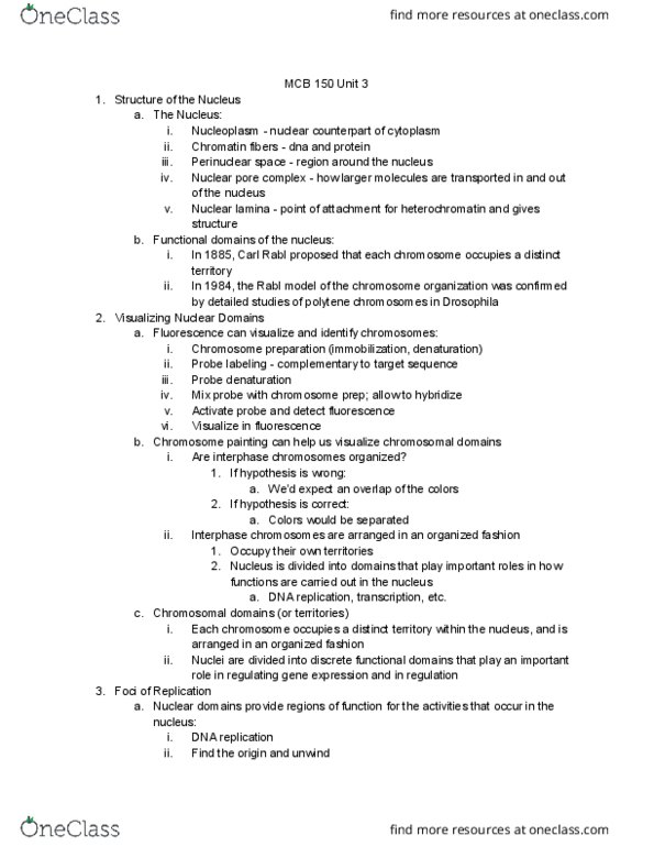
Nuclear envelope: double-lipid bilayer structure with outer and inner. Perinuclear space: space between inner and outer membrane. Nuclear pore complexes: how larger molecules are transported in and out. Nuclear lamina: point of attachment for heterochromatin, gives nucleus of nucleus shape and structure. Rabl hypothesis: each chromosome occupies distinct territory within interphase nucleus --> proven true by studies of polytene chromosomes in. These chromosomes are visible under light microscopy --> can now. Lends credibility that nucleus is more organized o o o. Visualizing nuclear domains o single strand dna bind to the dna. Denature remaining dna (no double strands) --> hybridize other molecules to. Probe labeling = molecule that binds is fluorescent and detectable --> can. Allow to hybridize with h bonds to single. Activate probe and detect fluorescence through fluorescence microscope stranded + complementary dna. Chromosomal domains (territories) chromosomes occupy and are organized in their own domains i. e. dna replication, protein ribosomal assembly, etc.