BIOL 4220 Lecture Notes - Lecture 8: Ferromagnetism, Positron, Positron Emission Tomography

Document Summary
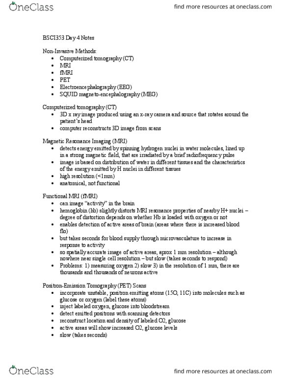
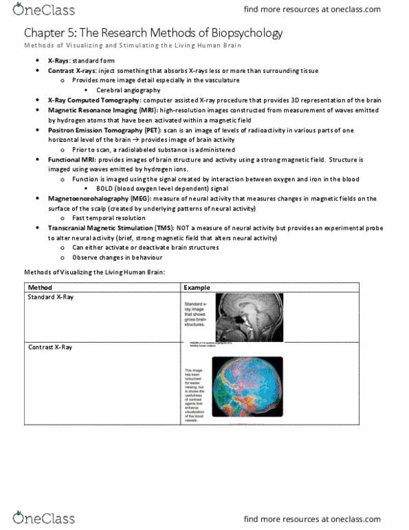
Positron emission as label for the radioactive tagged label. Ct scan: x-ray in 3d, use computer model to reconstruct 3d image, visualize the structure in 3d, x-ray measure hard tissue (radio-opaque) Pet scan (positron emission tomography: positron vs. electron, radioactive dye (molecule) tagged, emit positron (unstable) => neural firing => metabolic activity: corresponds to oxygen extraction, indirect measure of metabolism, measures bold (blood oxygen-level dependent) signal, measure brain activation. Dti (diffusive tensor imaging: measure axonal pathways, oscillate mri in magnetic field in specific directions, diffusion of water molecules fnirs (functional near-infrared spectroscopy, optical imaging, use near-infrared (nir) light shine onto brain. Diffusion is easier in longitudinal direction of axon. Diffusion is restricted in transverse direction of axon. Absorb with diff spectrum depending on oxy-hb or deoxy- Differentiate the diff btn oxy-hb and deoxy-hb conc. Measures similar to fmri, except can measure both oxy-hb and deoxy-hb.