Answer question part a-d
Question part A
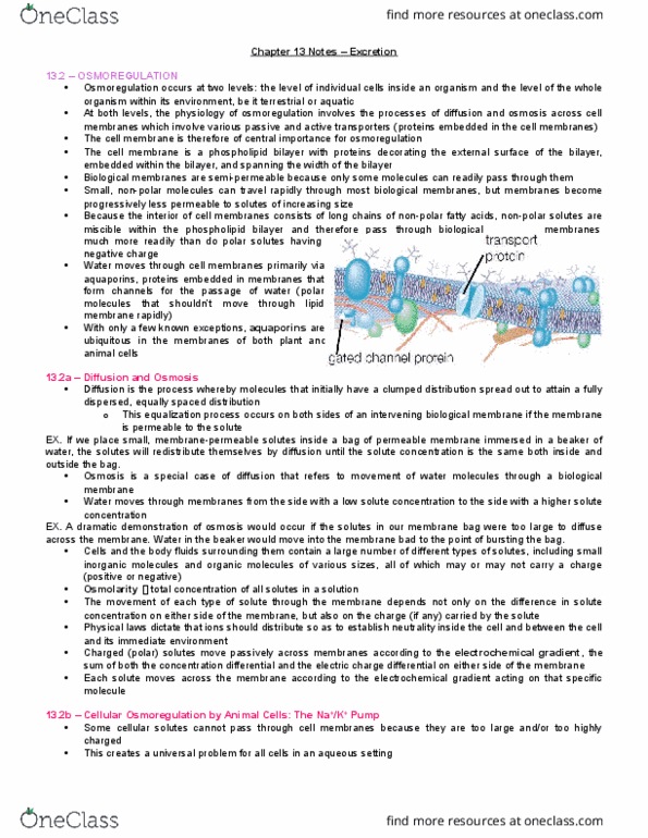
Diffusion Lab
Purpose: To investigate the diffusion of a substance across a semipermeable membrane.
Materials:
iodine (must be colored, clear won't work)
cornstarch/water solution
plastic sandwich bags (cheap is good)
beakers or clear plastic cups
Set-Up and Procedure
1. Fill beaker about halfway and put about ten drops of iodine.
2. Put about a teaspoon of starch and about 3 ounces of water in baggie and tie.
3. Make sure that it is water tight
4. Put the baggie in the beaker or glass and wait.
5. In about 15 minutes the solution in the baggie will turn blue/black. (purple)
This is a result of the iodine diffusing through the bag.
You should know that iodine is an indicator for starch. It turns blue/black in color.
Test it by dropping some iodine on a piece of white paper.
Questions and Conclusions :
Define permeability
To which substance is the plastic bag is permeable ?
Why did the iodine enter the bag ?
Why didn't the starch enter the beaker?
How is the plastic bag like the cell membrane?
Question part b
Reflex Lab
Procedures for Reflex Lab
In this lab we will test muscle stretch reflexes and cutaneous reflexes.
A partner is needed to be the subject for this lab.
Purpose: To test and observe normal muscle stretch and cutaneous reflexes.
Equipment: Reflex hammer,
Background: In the most narrow sense, a reflex is an involuntary, unpremeditated response by the body to an external stimulus. Reflexes allow reaction to a stimulus more quickly than would be possible if conscious thought were involved. A reflex can be described as a stimulus-response cycle. Most reflexes involve the brainstem or the spinal cord and do not require interaction from higher brain centers. The muscle stretch reflexes are the simplest reflexes. They involve the sensory neuron synapsing directly upon the motor neuron. They are sometimes called monosynaptic reflexes. They are also called spinal reflexes because the reflexive action does not require input from the brain. The other reflexes we will be testing in this lab are the cutaneous reflexes. They are also called superficial reflexes because the receptors are in the superficial areas of the body. They are more complex and require the message to go up the spinal cord to the brain and then back down to elicit the response. These can be used to test for CNS injury.
There are five components to a reflex; these components together are called the reflex arc. The components are as follows:
sensory receptor â receives the stimulus and generates an impulse (is usually a nerve ending)
afferent pathway â a neuron that carries the impulse from the receptor to a central integrating center
integrating center âthis is where the afferent component connects to the efferent component- this is in the CNS. One or more synapses must occur here. Interneurons are not involved in stretch reflexes but are always found in all other types of reflexes.
efferent pathway - a neuron that carries the impulse from the integrating center to an effector.
effector -receives the impulse and responds. The effector may by skeletal muscle, smooth muscle, or glands.
For more information please read chapter 12 : "Muscle" ( Pages 380-390) in Human Physiology book ( 12th edition by Stuart Ira Fox)
Part I. Stretch reflexes.
First do muscle stretch reflexes which are also called deep tendon reflexes.
It is important the subject relax the muscle being tested. Tell the subject to let the muscle go limp.
Rate each response according to the following scale:
+4 very brisk, hyperactive, large movement at joint.
+3 brisker than average, medium movement at joint
+2 average, small movement at joint
+1 somewhat diminished, very small or no movement at joint
0 no response
A. Patellar (also called the knee jerk reflex) â activation of femoral nerve. The receptor is located in the quadriceps muscle, the effector is the quadriceps muscle. The nerve ending is responding to the stretch of the muscle caused by striking the tendon.
1. A subject is allowed to sit comfortably on a table with knees dangling over the edge.
2. Using a reflex hammer (or substitute as described above) strike the patellar tendon just below the kneecap.
3. Observe the action caused by the reflex.
4. Record your results using the graded scale above.
B. Biceps Reflex â activation of the musculocutaneous nerve
1. Rest subjectâs left forearm along your left forearm.
2. Subjectâs hand should rest in or near the bend of your elbow.
3. Press your left thumb on the subjectâs biceps tendon, apply a little pressure.
4. Strike your thumb with the reflex hammer, this will transfer the pressure to the biceps tendon.
5. Watch carefully for twitching of the biceps muscle. Usually, the forearm will not move.
6. Record your results using the graded scale above.
C. Triceps reflex â activation of the radial nerve.
This is a difficult reflex to observe, but give it a try.
1. The subject should have the arm relaxed and elbow bent.
2. Support the entire weight of the arm with your left hand.
3. Strike the triceps tendons, just above the elbow.
4. Watch carefully for twitching of the biceps muscle.
5. Record your results using the graded scale above.
Both B and C show up best on someone who doesnât have a lot of subcutaneous fat and who has good muscle definition.
D. Achilles reflex (also called ankle-jerk reflex) â activation of the poplitealnerve. The principle is similar to the knee-jerk reflex. Here the stretch receptors are located in the gastrocnemius muscle.
1. Have the subject take her/his shoes and socks off, exposing the Achilles tendon on the back of the ankle.
2. Strike the Achilles with the reflex hammer (or substitute) and observe the extension of the foot downward.
3. Record the results using the graded scale above.
Part II. Cutaneous reflexes
A. Plantar reflex
This reflex differs from the four muscle stretch reflexes. In this reflex, receptors are stimulated in the sole of the foot (cutaneous receptors). The reflex pathway travels up the leg to the integrating center. While in the integrating center, the reflex travels up the spinal cord through the pyramidal tracts, which are nerve pathways between the spinal cord and brain.
1. The subject needs to lie on his or her back, with shoes and socks off.
2. Using the end of the handle of the reflex hammer, trace a path along the lateral side from heel to the little toe to the big toe.
3. Observe for the flexion (toe curling) which is recorded as a normal plantar reflex.
4. If the toes curl, this is called a Negative Babinski response and is normal. If the toes flare, then an abnormal response is recorded and is called a Positive Babinski response and indicates CNS damage.
5. Record the results.
B. Ciliospinal reflex â This reflexive pathway is from the skin to the thoracic spinal cord, through the sympathetic trunk, then to the dilator pupillae muscle of the eye. This is another test of the CNS.
1. Pinch the back of the subjectâs neck,
2. Watch pupils for a response.
3. A normal response is dilation of the pupil.
4. Record your results.
question part c
Homeostasis Lab - Heart Beat Regulation
This lab is designed to help explore the process of homeostasis as it applies to regulating heart rate. It also should help how a sampling process works.
Make two copies of the data sheet (or make your own) and paste them into your report.
1. Take a pulse for six seconds, record this number in the table below.
2. Every 15 seconds for the next three minutes, take a pulse again for six seconds, record each of these values.
3. Multiply each of these values by 10 to get the beats per minute.
4. Now average these numbers (this is the setpoint for the pulse at resting). Note the range of values (that is how far they can deviate from the set point).
5. The next step is to take a 30 second reading. Multiply this number by 2 to get beats per minute.
6. How does this compare to the setpoint?
7. Repeat steps 1 through 6 after running in place for 30 seconds. Record the same data.
8. After getting 8 readings, stop and plot these numbers on a piece of graph paper. The X-axis should be minutes and the Y-axis should be heart rate.(Don't forget to connect the points ). You can do this in Excel and then import it into Word. Or you can e-mail this to me.
Submit your lab report in the correct format.. Respond to the questions above. Explain WHY the results were what they were. Compare the resting and post-exercise and postulate explanations for the differences. You should have two data tables and one graph with your heart rate data from both tables on it in your report.
Time in seconds
Heart rate (bpm)
0 (initial recording)
15
30
45
60
75
90
105
question part d
What was one thing you found interesting, confusing or surprising about any of the lab activities? Why?
Answer question part a-d
Question part A
Diffusion Lab
Purpose: To investigate the diffusion of a substance across a semipermeable membrane.
Materials:
iodine (must be colored, clear won't work)
cornstarch/water solution
plastic sandwich bags (cheap is good)
beakers or clear plastic cups
Set-Up and Procedure
1. Fill beaker about halfway and put about ten drops of iodine.
2. Put about a teaspoon of starch and about 3 ounces of water in baggie and tie.
3. Make sure that it is water tight
4. Put the baggie in the beaker or glass and wait.
5. In about 15 minutes the solution in the baggie will turn blue/black. (purple)
This is a result of the iodine diffusing through the bag.
You should know that iodine is an indicator for starch. It turns blue/black in color.
Test it by dropping some iodine on a piece of white paper.
Questions and Conclusions :
Define permeability
To which substance is the plastic bag is permeable ?
Why did the iodine enter the bag ?
Why didn't the starch enter the beaker?
How is the plastic bag like the cell membrane?
Question part b
Reflex Lab
Procedures for Reflex Lab
In this lab we will test muscle stretch reflexes and cutaneous reflexes.
A partner is needed to be the subject for this lab.
Purpose: To test and observe normal muscle stretch and cutaneous reflexes.
Equipment: Reflex hammer,
Background: In the most narrow sense, a reflex is an involuntary, unpremeditated response by the body to an external stimulus. Reflexes allow reaction to a stimulus more quickly than would be possible if conscious thought were involved. A reflex can be described as a stimulus-response cycle. Most reflexes involve the brainstem or the spinal cord and do not require interaction from higher brain centers. The muscle stretch reflexes are the simplest reflexes. They involve the sensory neuron synapsing directly upon the motor neuron. They are sometimes called monosynaptic reflexes. They are also called spinal reflexes because the reflexive action does not require input from the brain. The other reflexes we will be testing in this lab are the cutaneous reflexes. They are also called superficial reflexes because the receptors are in the superficial areas of the body. They are more complex and require the message to go up the spinal cord to the brain and then back down to elicit the response. These can be used to test for CNS injury.
There are five components to a reflex; these components together are called the reflex arc. The components are as follows:
sensory receptor â receives the stimulus and generates an impulse (is usually a nerve ending)
afferent pathway â a neuron that carries the impulse from the receptor to a central integrating center
integrating center âthis is where the afferent component connects to the efferent component- this is in the CNS. One or more synapses must occur here. Interneurons are not involved in stretch reflexes but are always found in all other types of reflexes.
efferent pathway - a neuron that carries the impulse from the integrating center to an effector.
effector -receives the impulse and responds. The effector may by skeletal muscle, smooth muscle, or glands.
For more information please read chapter 12 : "Muscle" ( Pages 380-390) in Human Physiology book ( 12th edition by Stuart Ira Fox)
Part I. Stretch reflexes.
First do muscle stretch reflexes which are also called deep tendon reflexes.
It is important the subject relax the muscle being tested. Tell the subject to let the muscle go limp.
Rate each response according to the following scale:
+4 very brisk, hyperactive, large movement at joint.
+3 brisker than average, medium movement at joint
+2 average, small movement at joint
+1 somewhat diminished, very small or no movement at joint
0 no response
A. Patellar (also called the knee jerk reflex) â activation of femoral nerve. The receptor is located in the quadriceps muscle, the effector is the quadriceps muscle. The nerve ending is responding to the stretch of the muscle caused by striking the tendon.
1. A subject is allowed to sit comfortably on a table with knees dangling over the edge.
2. Using a reflex hammer (or substitute as described above) strike the patellar tendon just below the kneecap.
3. Observe the action caused by the reflex.
4. Record your results using the graded scale above.
B. Biceps Reflex â activation of the musculocutaneous nerve
1. Rest subjectâs left forearm along your left forearm.
2. Subjectâs hand should rest in or near the bend of your elbow.
3. Press your left thumb on the subjectâs biceps tendon, apply a little pressure.
4. Strike your thumb with the reflex hammer, this will transfer the pressure to the biceps tendon.
5. Watch carefully for twitching of the biceps muscle. Usually, the forearm will not move.
6. Record your results using the graded scale above.
C. Triceps reflex â activation of the radial nerve.
This is a difficult reflex to observe, but give it a try.
1. The subject should have the arm relaxed and elbow bent.
2. Support the entire weight of the arm with your left hand.
3. Strike the triceps tendons, just above the elbow.
4. Watch carefully for twitching of the biceps muscle.
5. Record your results using the graded scale above.
Both B and C show up best on someone who doesnât have a lot of subcutaneous fat and who has good muscle definition.
D. Achilles reflex (also called ankle-jerk reflex) â activation of the poplitealnerve. The principle is similar to the knee-jerk reflex. Here the stretch receptors are located in the gastrocnemius muscle.
1. Have the subject take her/his shoes and socks off, exposing the Achilles tendon on the back of the ankle.
2. Strike the Achilles with the reflex hammer (or substitute) and observe the extension of the foot downward.
3. Record the results using the graded scale above.
Part II. Cutaneous reflexes
A. Plantar reflex
This reflex differs from the four muscle stretch reflexes. In this reflex, receptors are stimulated in the sole of the foot (cutaneous receptors). The reflex pathway travels up the leg to the integrating center. While in the integrating center, the reflex travels up the spinal cord through the pyramidal tracts, which are nerve pathways between the spinal cord and brain.
1. The subject needs to lie on his or her back, with shoes and socks off.
2. Using the end of the handle of the reflex hammer, trace a path along the lateral side from heel to the little toe to the big toe.
3. Observe for the flexion (toe curling) which is recorded as a normal plantar reflex.
4. If the toes curl, this is called a Negative Babinski response and is normal. If the toes flare, then an abnormal response is recorded and is called a Positive Babinski response and indicates CNS damage.
5. Record the results.
B. Ciliospinal reflex â This reflexive pathway is from the skin to the thoracic spinal cord, through the sympathetic trunk, then to the dilator pupillae muscle of the eye. This is another test of the CNS.
1. Pinch the back of the subjectâs neck,
2. Watch pupils for a response.
3. A normal response is dilation of the pupil.
4. Record your results.
question part c
Homeostasis Lab - Heart Beat Regulation
This lab is designed to help explore the process of homeostasis as it applies to regulating heart rate. It also should help how a sampling process works.
Make two copies of the data sheet (or make your own) and paste them into your report.
1. Take a pulse for six seconds, record this number in the table below.
2. Every 15 seconds for the next three minutes, take a pulse again for six seconds, record each of these values.
3. Multiply each of these values by 10 to get the beats per minute.
4. Now average these numbers (this is the setpoint for the pulse at resting). Note the range of values (that is how far they can deviate from the set point).
5. The next step is to take a 30 second reading. Multiply this number by 2 to get beats per minute.
6. How does this compare to the setpoint?
7. Repeat steps 1 through 6 after running in place for 30 seconds. Record the same data.
8. After getting 8 readings, stop and plot these numbers on a piece of graph paper. The X-axis should be minutes and the Y-axis should be heart rate.(Don't forget to connect the points ). You can do this in Excel and then import it into Word. Or you can e-mail this to me.
Submit your lab report in the correct format.. Respond to the questions above. Explain WHY the results were what they were. Compare the resting and post-exercise and postulate explanations for the differences. You should have two data tables and one graph with your heart rate data from both tables on it in your report.
| Time in seconds | Heart rate (bpm) |
| 0 (initial recording) | |
| 15 | |
| 30 | |
| 45 | |
| 60 | |
| 75 | |
| 90 | |
| 105 |
question part d
What was one thing you found interesting, confusing or surprising about any of the lab activities? Why?