PSL201Y1 Lecture Notes - Pulmonary Vein, Pulmonary Artery, Thoracic Cavity

16 Oct 2011
School
Department
Course
Professor
Document Summary
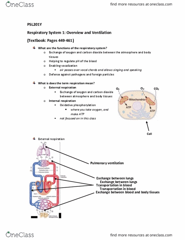
Allows for exchange of gases b/w air and blood. Defense from inhaled pathogens and foreign particles. Epiglottis prevents food from entering the lungs, vocal cords also close. Diaphragm contracts allow for lungs to expand during inspiration. Intercostal muscles raises ribcage to increase lung capacity. Internal intercostals contract to draw ribcage closer, reducing thoracic cavity: abdominal muscles contract to reduce volume. Pleural sacs (pleural fluid within pleural membranes enclose the lungs: allows for lungs to remain inflated against the walls of the thoracic cavity. Air is fully humidified and warmed to body temperature by arrival at the lungs. 1st division produces left and right main bronchi. 2nd-4th division produces lobar bronchi: have cartilage to maintain shape, cartilage ensures it remains open in presence of pressure. 12th-16th division produces terminal bronchioles, stabilized by bronchiolar muscles. No gas exchange occurs in all of these conducting airways, forming anatomical dead space. Between breaths of air, stale air is breathed in first.