Anatomy and Cell Biology 3319 Lecture : Anatomy Notes

1 May 2012
School
Department
Professor
Document Summary
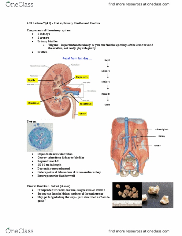
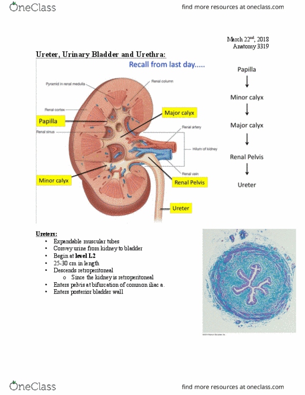
The ureters are muscular tubes that carry urine from the kidneys to the bladder. They stretch when they become filled and this creates a peristalsis-like movement that moves the urine down. They begin at the level of l2 (which is where the hilum is) as a continuation of the renal pelvis. They descend retroperitoneal (are not located in the peritoneal cavity). They enter the pelvis at the bifurcation of the common iliac artery and then enter the posterior bladder wall. The walls of the ureters have three layers a mucosa, a muscularis, and an adventitia. There are three locations at risk for kidney stones the renal pelvis/proximal ureter, the pelvic brim/common iliac, and the wall of the bladder. Urinary bladder: the bladder is a pyramid shaped muscular sac with a base, apex, and body. It lies inferior to the peritoneal cavity and posterior to the pubic symphysis.