ANHB2212 Study Guide - Final Guide: Vaginal Process, Celiac Plexus, Rectus Abdominis Muscle

3 Jun 2018
School
Course
Professor

Abdomen
Intra-Embryonic Coelom:
• Coelom → space that develops within the mesoderm
• Intra-embryonic coelom develops within intra-embryonic mesoderm
during week 3
• Divides lateral plate mesoderm into
o Somatopleure
o Visceropleure
• Derivatives of the intra-embryonic coelom are spaces separating the body
walls (somatopleure) from viscera (visceropleure)
• Derivatives of the intra-embryonic coelom
o Pericardial cavity
o Pleural cavity
o Peritoneal cavity
Development of Body Cavities:
• Organs are surrounded by cavities → do not lie in it
• Organs develop adjacent to the coelom than invaginate into cavity
• As they grow into the cavity they become coated by a layer of serous
membrane
o Visceral layer → lines organ surface
find more resources at oneclass.com
find more resources at oneclass.com

o Parietal layer → lines body cavity
o Serous caity → surrounds organ
• Connecting layer between visceral and parietal layers → mesenteries
• Development of serous cavities
o Begin as a horse-shoe cavity in embryonic disc
o Arch passes in front of oral membrane and cranial end of neural
tube → behind cardiogenic area and septum transversum
o As brain grows, embryo folds sagittally → septum transversum
and developing heart lie ventrally
o Coelom horseshoe folded to become 3 sections
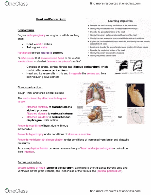
▪ Pericardium → heart in the middle
▪ Left and right pleuroperitoneal cavities
o Week 4 → pericardium is partitioned from rest of coelom
▪ Septum transversum developed up near head so receives
its nerve supply (phrenic nerve) from cervical region
▪ When folding occurs, phrenic nerves are dragged down
thorax
▪ Phrenic nerves come to lie between the pericardium and
pleuroperitoneal canals → help separate cavities
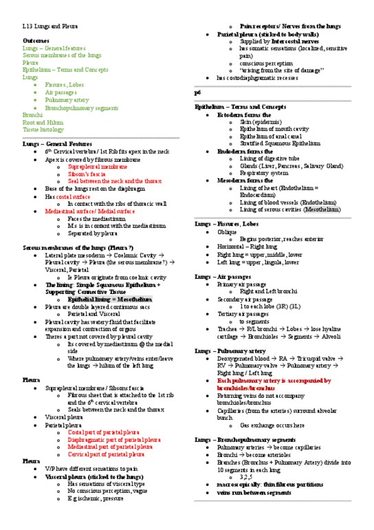
• Development of pleural cavity
o Foregut (osephagus) runs behind developing heart →
pleuroperitoneal canals on either side
o Foregut gives rise to lung buds that grow out laterally into
left/right pleuroperitoneal canals
o Diaphragm separates thorax from abdomen
o Pleuroperitoneal canals are partitioned by growth of diaphragm
▪ Septum transversum (centrally)
▪ Pleuralperitoneal membranes (posteriorly)
▪ Ingrowth of body wall (laterally)
o Sometimes pleuroperitoneal membranes fail to complete the
formation of the diapgragm → allows abdominal organs to
herniate into thorax
• Development of peritoneal cavity
o Mesenteries contain blood vessels that attach to posterior body
wall
o Initially peritoneal cavity is in left and right parts (canals)
▪ Gut → both dorsal and ventral mesenteries
▪ Dorsal mesentery → contains arteries from aorta
o Gut tube divided according to blood supply
▪ Foregut → coeliac artery
▪ Midgut → superior mesenteric artery
▪ Hindgut → inferior mesenteric artery
o Foregut
▪ Characterized by structures developing in dorsal and
ventral mesenteries → mesogastrium
▪ Ventral mesogastrium
• Liver
• Ventral pancreas
▪ Dorsal mesogastrium
find more resources at oneclass.com
find more resources at oneclass.com

• Dorsal pancreas
• Spleen
o Midgut and hindgut
▪ Lose ventral mesentery
▪ Left and right canals merge to form a single cavity
▪ Midgut → characterized by a rapid growth in length
• Midgut hernia → returns to abdominal cavity by
week 10
• Rotation around axis of superior mesenteric artery
brings caecum and appendix to right
▪ Hindgut → characterized by partition of primitive cloaca
into rectum and bladder
• Urogenital septum → bladder in front, rectum
behind, genitals in between
The Abdominal Cavity:
• Functions
o Houses and protects abdominal viscera
o Plays a role in breathing → forced exhalation
o Control of intra-abdominal pressure
o Posture and core stabilization
• Abdominal regions → 9 zones → 3x3
o Top row → along costal margin → T8/9 (xiphisternal joint)
▪ Right and left hypochondrium
▪ Epigastric
o Middle row → L3/4 (umbilicus)
▪ Right and left lumbar
▪ Umbilical
o Lower → along pelvis → coccyx
▪ Right and left iliac
▪ Hypogastric
find more resources at oneclass.com
find more resources at oneclass.com