ANHB2212 Study Guide - Final Guide: Atrioventricular Canal, Sinus Venosus, Circumflex Branch Of Left Coronary Artery

3 Jun 2018
School
Course
Professor

The Heart
Pericardium:
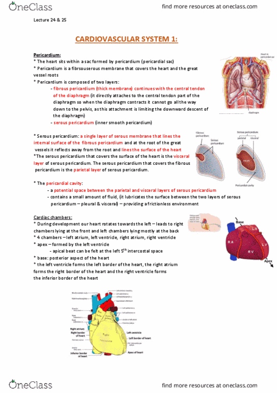
• Fibrous sac that surrounds the heart in the middle mediastinum
• Situated between pleural cavities
• Tough fibrous bag that holds the heart in position
• Consists of a strong, conical fibrous sac (fibrous pericardium), within
which is the serous pericardium
• Heart and its great vessels lie within this structure and invaginate the
serous sac from behind during development
• Fibrous pericardium
o Tough, thick, flask like sac
o Neck is closed by attachment to great vessels
o Attachments
▪ Ventrally → manubrium and xiphoid process
▪ Dorsally → vertebral column
▪ Caudally → central tendon
o Base attached to central tendon and diaphragm → helps hold heart
in position and limit movement
o Prevents
▪ Overfilling due to relative inextensible fibrous layer
▪ Hypertrophy of the heart under conditions of strenuous
exercise
▪ Ventriculo-atrial regurgitation under conditions of
increased ventricular end-diastolic pressures
o Acts as a physical barrier between muscular body of the heart and
adjacent organs → affords protection from infection
• Serous pericardium
o Covers outside of heart → visceral pericardium
o Lines the insides of the fibrous sac → parietal pericardium
o Transparent membrane forming a closed sac containing small
amount of lubricating fluid
o Essentially forms a bursa that facilitates cardiac movement
find more resources at oneclass.com
find more resources at oneclass.com

• Pericardial cavity
o Slender space between opposing visceral and parietal surfaces
o Normally contains between 10-20mL of pericardial fluid secreted
by pericardial membranes
o Acts as a lubricant that reduces friction between opposing
surfaces, also some degree of shock absorbance
o Moist pericardial lining also prevents friction as the heart beats
o This in combination with the collagen fibers anchoring the base of
the heart to the mediastinum limits movement of major vessels
• Vascular supply
o Fibrous and parietal
▪ Arterial supply
• Internal thoracic artery
• Descending thoracic aorta
▪ Venous drainage
• Azygous vein
• Internal thoracic vein
o Visceral
▪ Arterial supply
• Coronary arteries
▪ Venous drainage
• Coronary sinus
• Nerve supply
o Fibrous and parietal → phrenic nerve
o Visceral → vagus and sympathetic nerves
Sinuses:
• Transverse
o Lies behind the ascending aorta and main pulmonary artery
o Extends transversely between right and left pericardial spaces
o Communicates the two halves of the pericardial cavity
o Acts as a bursa → containing pericardial fluid between pulsating
arteries in front and the contrasting atria behind
• Oblique
o Passes upward and to the right behind the left atrium anteriorly
find more resources at oneclass.com
find more resources at oneclass.com

o Lies in front of the oseophagus and descending thoracic aorta
posteriorly
o Believed to act as a bursa for the left atrium to expand during
filling
Gross
Anatomy of the Heart:
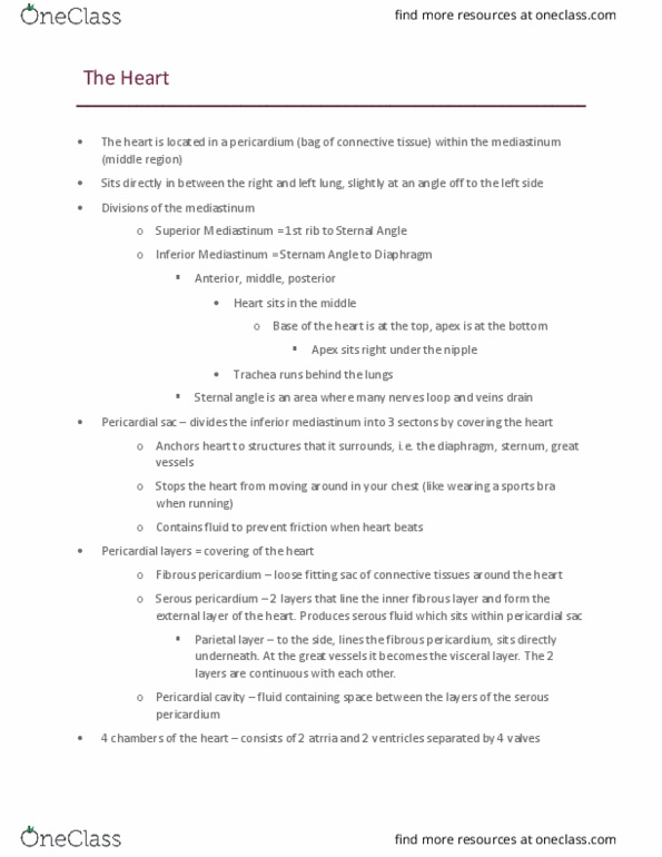
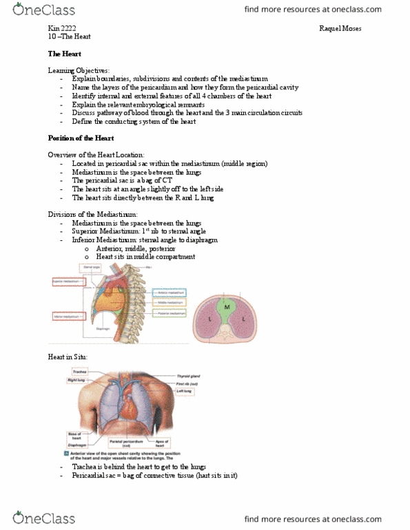
• Orientation
o Middle mediastinum
o Positioned slightly left to the midline
o Lies between sternum and vertebral column
o Sits at an oblique angle to longitudinal axis of body
▪ Base forms superior border → 3rd costal cartilage
▪ Right border formed by right atrium
▪ Left border formed by left ventricle and small portion of left
atria, extending to apex where it meets inferior border
▪ Inferior border formed mainly by inferior wall of left
ventricle
o Typical adult heart → 12.5cm from base to apex
o Apex usually reaches 5th intercostal space ~7.5cm left of midline
find more resources at oneclass.com
find more resources at oneclass.com