PHY2011 Study Guide - Final Guide: Fallopian Tube, Theca, Endometrium

6 Jun 2018
School
Department
Course
Professor

Lecture 3: Female Reproduction 2
Lecture Objectives
1. Describe the structure and function of the female reproductive organs
2. Explain the complete ovarian and uterine cycles
3. Understand the hormonal mechanisms that regulate female reproductive functions
Endocrine system & HPG axis
• Hypothalamus produces GnRH
o Gonatrophin Releasing Hormone aka: luteinising hormone-releasing hormone (LHRH)
o Controls the reproductive cycle
o 10 amino acid peptide (composed of amino acids) hormone / hypophysiotrophic (acting on the
pituitary gland) hormone
o Released from 1000’s neurons within the hypothalamus in a pulsatile (not continuous) manner
§ Produced in neurones in the brain (pre-optic area and mediobasal hypothalamus)
§ GnRH neurones secrete GnRH into hypophysial portal system in pulses
o The GnRH pulse generator with 1⁄2 life of GnRH is minutes à has to keep a certain amount of
GnRH to be released
o GnRH pulses stimulate the synthesis and secretion of LH and FSH from the anterior pituitary
• Pituitary Gland produces LH & FSH
o Gonadotrophins: Luteinising Hormone (LH) and Follicle Stimulating Hormone (FSH)
o Glycoprotein hormone (long chains of amino acids)
o Heterodimeric polypeptides (α- and β-subunit linked by disulfide bonds)
§ α-subunit is common to both LH and FSH (and hCG)
§ β-subunit is unique and confers biological specificity
o Gonadotrophins are synthesised and stored in gonadotroph cells in the anterior pituitary
o Gonadotrophins are secreted into the peripheral circulation
o LH secretion is pulsatile
o FSH secretion is not pulsatile (passive)
Gonadotrophins act on the gonads to stimulate the production of the gametes (sperm in males and oocytes in
females) and to stimulate the production of gonadal hormones such as the sex steroids

• Ovaries produce estrogens, progesterone and inhibin (all families of hormones)
Sex steroids (Androgens, estrogens and progestogens)
• Steroid hormones (common characteristics à lipid soluble; lipophilic, enables them to diffuse through lipid
bilayer)
• Derived from cholesterol à synthesized by cholesterol
• 4 ring structure
In females, androgens are produced in the ovary by thecal cells (cells which form outside of a developing follicle
and develop androgens)
• Androgens are converted to estrogens in the ovary by granulosa cells
• The corpus luteum of the ovary produces progesterone
• Sex steroids are also produced by the cortex of the adrenal gland
• Sex steroids have actions within the ovary and are secreted into the peripheral circulation
• Important roles in a range of reproductive functions including production of the gametes and feedback
actions on the reproductive axis (HPG axis)
In females estrogens exerts negative/positive feedback effects; Progesterone exerts only negative feedback effects
Inhibin
• Glycoprotein hormone
• In females, inhibin is produced in the ovary by granulosa
cells (inhibin A and B)
• The corpus luteum also produces inhibin
• Inhibin is secreted into the peripheral circulation
• Feedback actions on the reproductive (HPG) axis
• In particular, negative feedback actions on FSH secretion by
direct actions at the anterior pituitary only
o Doesn’t influence hypothalamus
• Eg; Inhibin B (produced in the testes)

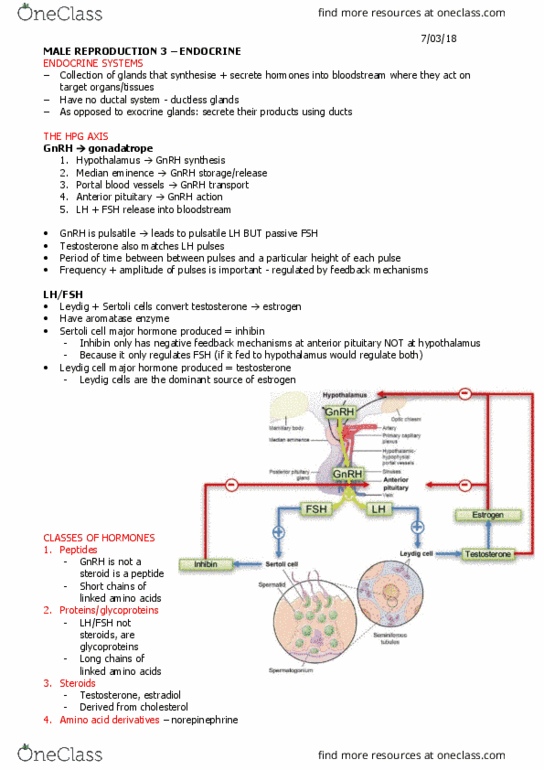
The hypothalamo-pituitary axis
§ Hormone will have an effect on a target organ
§ That target organ will have the capacity to react to how
much that hormone is present
§ Too much à target organ has potential to alter the
amount present
§ If there is too much present, it can be reduced
§ If there is too little, it can be increased
o If the leydig cells produce too much
testosterone, hypothalamus signals LH to
reduce production levels
§ Hypothalamus serves as the link between the nervous
system and the endocrine system à achieves this
through close relation with the anterior pituitary gland
o GnRH travels down to median eminence (and is
stored and released there)
o Within vascular network where GnRH is released à has a short half-life within the blood so needs
to travel quickly
o Secretes LH & FSH which effects the Gonads
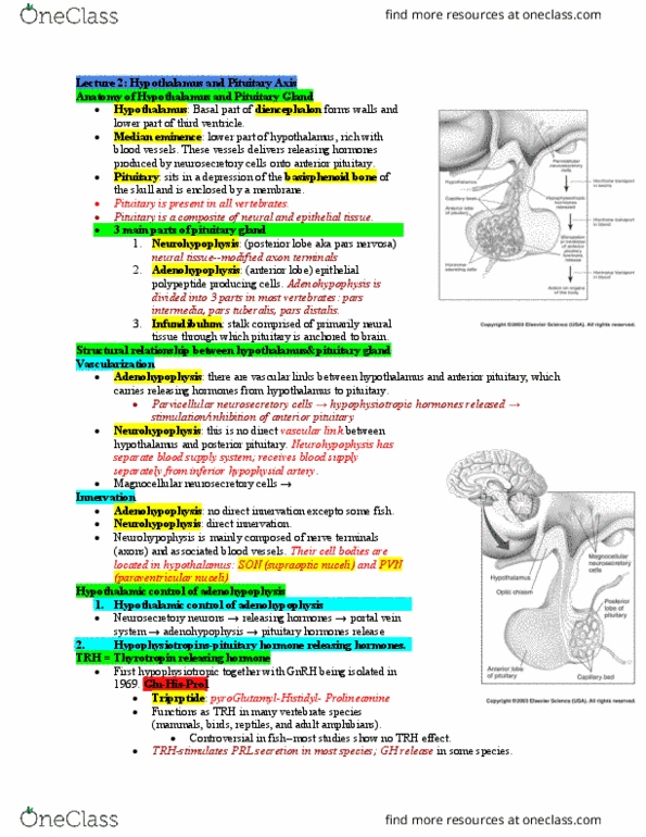
Pulsatile & passive release
Pulsatile
§ How hormones are released
§ GnRH – has a pulsatile release
§ Surge at regular intervals, so not released in 1 continuous
amount à released periodically
§ LH is released in response to each of the GnRH pulses
§ Both releases are pulsatile
Passive
§ FSH release is done in release to GnRH but not pulsatile
§ It has a passive release
§ Response to pulse frequency (number occurring over a period
of time) à If GnRH decreases, FSH levels drop
Document Summary
Lecture objectives: describe the structure and function of the female reproductive organs, explain the complete ovarian and uterine cycles, understand the hormonal mechanisms that regulate female reproductive functions. Produced in neurones in the brain (pre-optic area and mediobasal hypothalamus) Gnrh neurones secrete gnrh into hypophysial portal system in pulses: the gnrh pulse generator with 1 2 life of gnrh is minutes has to keep a certain amount of. Subunit is common to both lh and fsh (and hcg) Subunit is unique and confers biological specificity: gonadotrophins are synthesised and stored in gonadotroph cells in the anterior pituitary, gonadotrophins are secreted into the peripheral circulation, lh secretion is pulsatile, fsh secretion is not pulsatile (passive) Sex steroids (androgens, estrogens and progestogens: steroid hormones (common characteristics lipid soluble; lipophilic, enables them to diffuse through lipid bilayer, derived from cholesterol synthesized by cholesterol.