HTHSCI 1DT3 Study Guide - Midterm Guide: Cx3Cl1, Morphogen, Wnt1

23 Jun 2018
School
Department
Course
Professor

(SGZ) of hippocampus
SVZ cells are glial lineage (GFAP+ve) ‘Type B’ Cells, and unlike in embryonic
stage they are quiescent in adults. Can divide asymmetrically (maintain stem cell
pool).
Type B Cells can divide into Type C Cells that form Type A cells which are
neuroblasts, that can migrate to olfactory bulb (via rostral migratory stream) in rats –
which rely heavily on sense of smell.
Song et al (2012) showed how spill over GABA from surrounding neurons in SGZ
can stop stem cell proliferation (leaky synapses?). Leakage of GABA keeps stem
cells in quiescence.
Disorders of Cortical Development
Failure of proliferation – microcephaly
Failure of neuronal migration – periventricular heterotopia
Overmigration of neurons to pial surface – cobblestone lissencephaly
Reeler mouse mutants – gross malpositioning of neurons in cerebral and cerebellar cortex:
In cerebral cortex neurons fail to migrate past ‘older’ neuronal layers, and form
‘outside in’ development of cerebral cortex – wrong.
In cerebellum, reduced granule cell number and Purkinje Cells aggregate instead of
forming a monolayer.
Conclusion
Misc Notes:
Reelin binds to ApoER2 and VLDLR receptors on migrating neuroblasts, causing
downstream activation of Dab1.
Currently unsure whether Reelin acts as a ‘stop’ or ‘go’ signal, with evidence
suggesting and disproving both theories. Possible that Reelin may act as ‘go’ signal
during neuronal migration, and ‘stop’ signal once cells have reached correct cortical
level.
Studies have shown that increasing Dab1 degradation (i.e. reduced Reelin effects) causes
postmitotic neurones to fail to migrate past previous layer, while reduced Dab1 degradation
cause overmigration
•
•
•
•
o
o
o
o
•
•
•
•
•
•
o

The development, survival, maintenance and remodelling of neurons depends on the cytoskeleton.
Discuss.
Alzheimer’s Disease (Theory, biochemistry, APP, NGF) with relationship to transport failure?
Discuss the role of the neuronal cytoskeleton in intracellular transport, and how this is important in
neurogenesis and development.
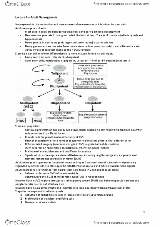
Introduction
Cytoskeleton plays important role in mechanical strength, morphological plasticity/cell
movement and intracellular transport.
Acts as a cellular scaffold that gives cells (including neurones) structure. Found in all cells,
and plays a vital role in strengthening and transporting components through neuronal
axons and dendrites.
Cytoskeleton split into three protein components:
Microfilaments (smallest, actin, involved in morphological plasticity),
Intermediate filaments (variable, GFAP – astrocytes, neurofilament in neurones,
provide strength)
Microtubules (largest, organised physical cylinders, involved in cellular transport)
Particularly important during neurodevelopment – neurones rely on cytoskeleton for
movement and pathfinding (immature neurons have growth cones that later develop into
presynaptic knobs – rely on cytoskeleton to dynamically adjust growth cone movement).
Response to inductive and inhibitory cues allows for dynamic changes in cytoskeleton to
allow for precise control of neuronal development in CNS.
Actin filaments – Morphological Plasticity
Actin filaments formed from actin subunits. Dynamic changes to actin filaments allow for
movement of growth cone during neurodevelopment, in response to particular cues that
can be attractive or repulsive.
Dynamic nature stems from Arp Complex and action of two key proteins: profilin and
cofilin. Profilin adds filaments to plus end and allows filament elongation, whilst cofilin is
involved with breaking down actin filaments to ‘free’ up available actin subunits to be used
at plus end (leading edge/growth cone).
Cofilin activity inactivated by phosphorylation (LIM Kinase), and reactivated by
dephosphorylation (slingshot).
Combination of profilin and cofilin allows plasticity changes in growth cone via actin
filaments, with support from microtubule that provides additional stability.
Actin cytoskeleton can also be influenced by Rho GTPases (from Ras molecular switch
family). RhoA stimulates stress fibres formation (involved in growth cone collapse), Rac1
stimulates Lamellipodia formation, Cdc42 stimulates fillopodia formation
(Rac1 + Cdc42 involved in growth cone advance and axonal growth).
Actin filaments also key in synapse function in synaptic plasticity (both during
development and adult?) – involved in both presynapse (controls RESERVE POOL of
synaptic vesicles, and docking of READY-RELEASABLE POOL).
In postsynaptic membrane, actin meshworks hold protein and receptors in place and
control shape of spine.
Synaptic strength (and degree of plasticity?) associated with degree of actin polymerisation
in dendritic spines (postsynaptic membrane) influenced by Arp complex.
Intermediate filaments – Axonal Strength
IF variable depending on cell type, with functions relevant to lineage.
GFAP in astrocytes, neurofilaments in neurones.
NF have high tensile strength, and providing good support especially given small diameter
and long lengths that axons can reach.
NF bend easily, difficult to break – providing support and enhancing survival of neurone.
Especially given the fact they need to last a lifetime (postmitotic).
Microtubules – intracellular transport
Largest protein component of cytoskeleton, important in both developing neurons
(extending axons towards target tissues) and mature neurons (with established synaptic
connections).
Differences in MT between dendrites and axons.
MT stabilised by additional proteins (Tau in neurones, MAP2B in dendrites).
Axons – MT arranged in uniform direction (same polarity) facing plus end
(towards growth cone/synapse), forming a transport track.
•
o
o
o
•
•
•
o
o
•
o
o
o
o
o
o
o
o
•
o
o
o
o
•
o
o
•
•

Dendrites – disorganised layout with MT having mixed polarities.
MT Axonal function can be split into slow axonal transport and fast axonal transport.
Slow axonal transport – e.g. of ‘housekeeping’ components (actin, NF, MT,
organelles)
Dynein like motors carry actin cytoskeleton, while MT used to transport NF.
Fast axonal transport – has anterograde and retrograde transport, and make use of
cytoskeletal motor proteins.
Kinesins – move towards plus end (from soma to growth cone/synapse) –
contribute to ANTEROGRADE TRANSPORT (e.g. carry growth factor
receptors, mitochondria etc.)
Dyneins – move towards minus end (back to soma, from growth/cone
synapse) – contribute to RETROGRADE TRANSPORT (e.g. carry growth
factor signalling complexes to soma)
Vesicles bind to tail portions of kinesin/dynein motor proteins (via transmembrane
receptors) – one such transmembrane protein is APP, needed in fast anterograde
transport (kinesin).
Goldstein Group, and Kamal (2000) – Suggested APP was a key receptor important
for coupling vesicle to kinesin.
Later shown by Lavrov (2005) to not bind directly, but evidenced APP-kinesin
interaction still existed but perhaps through a more complex mechanism.
APP can be cleaved by three proteases near lipid bilayer of vesicle:
Alpha + Gamma secretase – causes release of large APP ectodomain, P3 peptide and
AICD (intracellular short C-terminal) – harmless
Beta + Gamma secretase – causes release of another large APP ectodomain, Aß
peptide and AICD. Aß peptide can form plaques.
Plaques consisting of Aß peptide have been identified as a key pathological finding
in Alzheimer’s disease and are extracellular insoluble deposits (along with
NFT/PHF/hyperphosphorylated tau).
Alzheimer’s disease – degeneration of neurones, especially frontal, temporoparietal
and hippocampal – leading to cognitive and short-term memory problems.
But do the Aß plaques themselves cause Alzheimer’s, or are they simply an
exacerbating byproduct of another disease process?
Possibility that Aß plaques could interfere with intracellular transport mechanisms?
Ekinci (2000) suggested that Aß plaques result in increased intracellular Ca2+ and
ROS – both leading to apoptotic pathway activation?
Zempel (2010) further suggested how failure of local elevation of intracellular
Ca2+ was associated with failures of microtubule transport of mitochondria and
raised intracellular ROS.
Isacson and colleagues (2002) suggested that APP could cause the disease, not Aß.
Possible theory that APP is responsible for carrying TrkA to presynapse (plus end).
TrkA receptor binds to NGF, a key neurotrophin responsible for neuronal
survival/growth.
Failure of TrkA transport by APP reduces NGF-TrkA mediated neuronal
survival/trophic support and death of neurone (observed in Alzheimer’s).
Conclusion
Above discussion suggests importance of cytoskeleton in neuronal function – both during
development and in mature CNS environment and key role in support/strength, plasticity
and intracellular transport.
Perhaps (as evidenced by Alzheimer’s theory) to play a role in key neurodegenerative
disease and may provide a possible clue to understanding disease pathways and possible
therapeutic interventions?
•
o
•
•
•
•
•
•
•
•
o
•
•
•
•
•
•
•
•
•
•
•
•
•
o
o
Document Summary
Svz cells are glial lineage (gfap+ve) type b" cells, and unlike in embryonic stage they are quiescent in adults. Type b cells can divide into type c cells that form type a cells which are neuroblasts, that can migrate to olfactory bulb (via rostral migratory stream) in rats which rely heavily on sense of smell. Song et al (2012) showed how spill over gaba from surrounding neurons in sgz can stop stem cell proliferation (leaky synapses?). Leakage of gaba keeps stem cells in quiescence. Disorders of cortical development o o o o. Overmigration of neurons to pial surface cobblestone lissencephaly. Reeler mouse mutants gross malpositioning of neurons in cerebral and cerebellar cortex: In cerebral cortex neurons fail to migrate past older" neuronal layers, and form. Outside in" development of cerebral cortex wrong. In cerebellum, reduced granule cell number and purkinje cells aggregate instead of forming a monolayer.