NSE 13A/B Study Guide - Midterm Guide: Sternal Angle, Thoracic Vertebrae, Costal Cartilage

26 Jan 2016
School
Department
Course
Professor

Document Summary
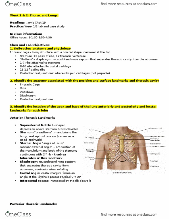
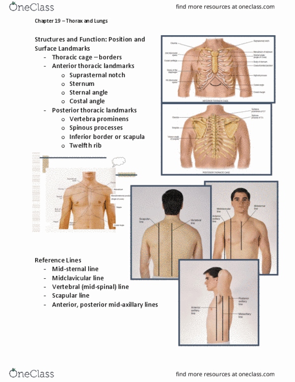
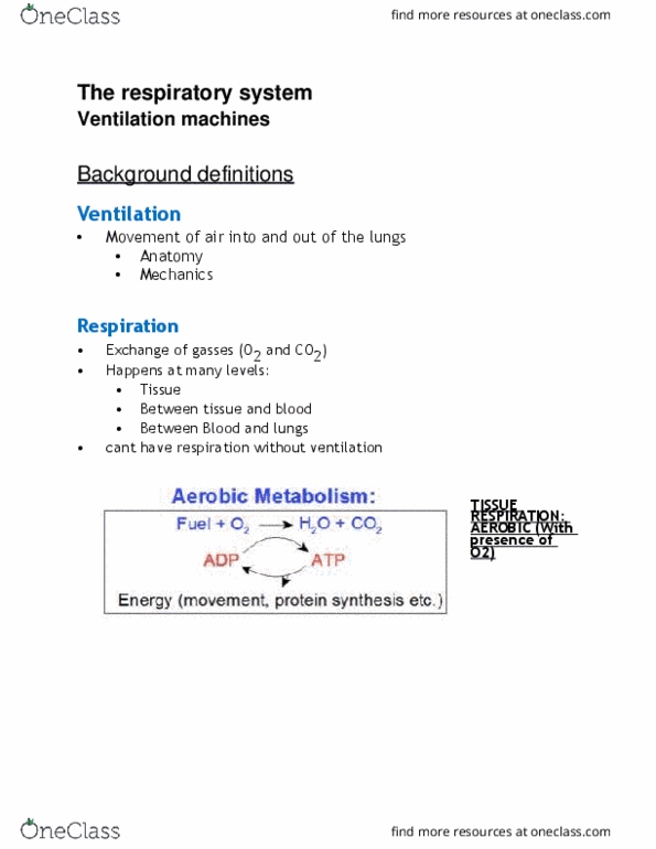
Nse 13b- assessment of healthy individual: week 5 objectives. Jarvis chapter 19: self-review anatomy and physiology. Normal stimulus to breathe is an increase in co2 in the blood or hypercapnia. A decrease in o2 (hypoxemia) will also increase respirations but not as effective as hypercapnia. Identify the anatomy associated with the position and surface landmarks and thoracic cavity. Thoracic cage is a bony structure with a conical shape, which is narrower at the top: defined by the sternum, 12 pairs of ribs, and 12 thoracic vertebrae. Its bottom is the diaphragm, separates the thoracic cavity from the abdomen. The anterior aspects of the first 7 ribs attach directly to the sternum via their costal cartilages; those of ribs 8,9, and 10 attach to the costal cartilage above; and those of ribs 11 and 12 are. The costochondral junctions are the points at which the ribs join their cartilages.