PAT 20A/B Study Guide - Final Guide: Extracellular Fluid, Intracerebral Hemorrhage, Diuretic

10 Mar 2017
School
Department
Course
Professor
Document Summary
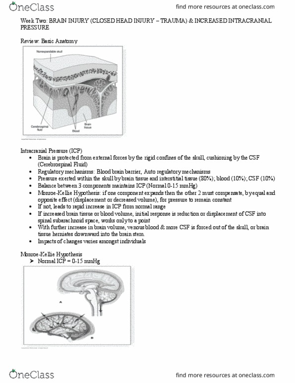
The adult brain is enclosed within a nonexpendable rigid skull or cranium making it susceptible to increases in icp. Excessive icp can: obstruct cerebral blood flow destroy brain cells ( as in herniation) and damage delicate brain structures. The skull is like a closed box with 3 essential components: cranial cavity. Monro- kellie hypothesis: normal fluctuations in icp occur with respiratory movements and adl such as straining, coughing, and sneezing. Volume change in any of the 3 compartments = abnormal variation in intracranial volume + changes in icp. Increase in tissue volume can result from: brain tumour cerebral edema bleeding into brain tissue. An increase in blood volume develops: vasodilation of cerebral vessels when there is obstruction of venous outflow. Changes in the cerebral spinal fluid compartment occurs from: Buffered by a translocation of csf to the spinal subarachnoid space and increased reabsorption of csf.