PHYSL212 Study Guide - Ciliary Muscle, Intraocular Pressure, Retina

13 Dec 2014
School
Department
Course
Professor

Document Summary
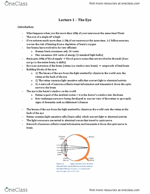
Components: optical focus the image on receptor cells, neural transforms visual image into action potentials. Optical component: sclera - protection, cornea allows light through. Lens responsible for focusing on moving object: ciliary muscle move lens. Refraction is a path of light coming from density a to density b. High density point is eye (inside) light bends inwards. Primary structure responsible for refraction is cornea. Long distance: ciliary muscle relaxed, flat lens: close distance, out of focus: relaxed ciliary muscle, in focus: ciliary muscle contracted, lens rounded. Presbyopia loss of elasticity of the lens, results in inability to accommodate for near vision. Myopia too long eyeball, image doesn"t reach the back of the eye; concave lens correction. Hyperopia too short eyeball, image focuses behind focal point; convex lens correction. Astigmatism surface of lens/cornea isn"t smoothly spherical; surgery. Glaucoma damage of retina due to increased intraocular pressure (too much vitreous humor) Rods photoreceptors for low light conditions (grey).