ANP 1106 Study Guide - Midterm Guide: Dorsal Root Ganglion, Filum Terminale, Lumbar Puncture

19 Apr 2016
School
Department
Course
Professor
Document Summary
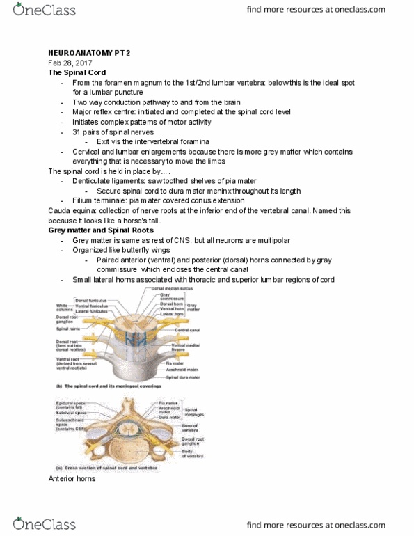
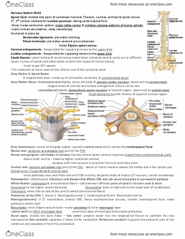
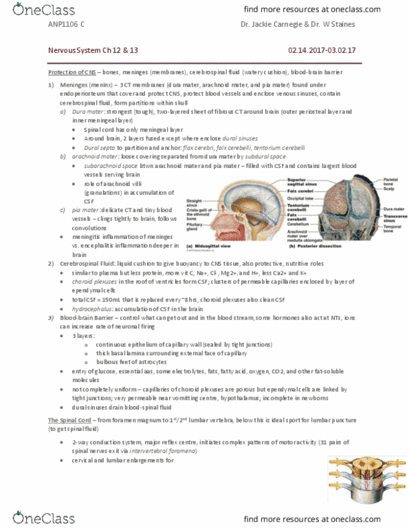
From foramen magnum to 1st/2nd lumbar vertebra, below this is ideal spot for lumbar puncture: 2 way conduction system, major reflex centre, initiates complex patterns of motor activity. Cervical and lumbar enlargements where the nerves serving the lower and upper limbs arise. Spinal cord held in place by: denticulate ligaments (anchor pia to dura, filum terminale (anchor for spinal cord) Cauda equine: collection of nerve roots at the inferior end of the vertebral canal. Ventral and dorsal horns connected by gray commissure (encloses central canal) Nerve cell bodies of somatic motor neurons axons exit via ventral roots. Largest at levels of cervical and lumbar enlargements. Sympathetic motor neurons to visceral organs, also exit via ventral roos. Funiculus: tracts or neurons, have specific origin and destination. Myelinated and unmyelinated nerve fibers that communicate between different parts of cord and between cord and brain. Fibers run in 3 directions: ascending, descending and transverse (commissural) tracts. Made of ventral and dorsal spinocerebellat tracts.