Anatomy and Cell Biology 3309 Study Guide - Final Guide: Cumulus Oophorus, Anterior Chamber Of Eyeball, Posterior Chamber Of Eyeball

22 May 2018
School
Department
Professor

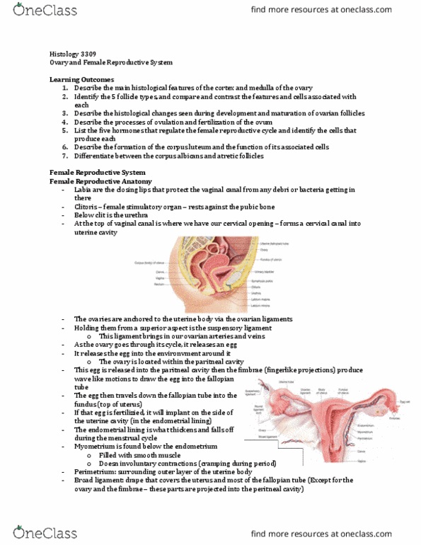
Histology 3309
Lab 24
Eye
Identify the outlined structure
- Unilaminar primary follicle
o Unilamilar bc only 1 layer of follicular cells
- The oocyte is in the middle but when the cells are incorporated, its some sort of follicle
Identify the outlined structure
- Graafian follicle
Identify the outlined structure
- Yellow outline = where the oocyte would be
- No antrum so its not secondary
- It’s a multilaminar primary follicle
Identify the outlined structure
- Cumulus oophorus →connection within the Graafian follicle to the oocyte
find more resources at oneclass.com
find more resources at oneclass.com

Identify the layer outlined by the box
- Stratum basalis of the endometrium
- Lower third of the endometrium
- More basophilic
- The coils of the coiled glands will be in the functionalis
Eye
- A = anterior
- P = posterior
- Blue bracket = cornea
o Pale staining layer where air is going to hit
o Most anterior layer
- AC = anterior chamber (aka aqueous chamber)
o Space that is just posterior to the cornea
o Find aquous humour (aka aquous fluid) here
- Black bracket = lens (L)
o In most slides, its going to be torn
- Green bracket = vitreous chamber (VC)
o Have vitreous humor here – gelly like fluid
- BS = blind spot
- ON = optic nerve
- There are musle attachments to allow us to move the eye (on the side black circles)
- Retina (R ) (blue bracket)
find more resources at oneclass.com
find more resources at oneclass.com
Document Summary
Unilaminar primary follicle: unilamilar bc only 1 layer of follicular cells. The oocyte is in the middle but when the cells are incorporated, its some sort of follicle. Yellow outline = where the oocyte would be. Cumulus oophorus connection within the graafian follicle to the oocyte. The coils of the coiled glands will be in the functionalis. Blue bracket = cornea: pale staining layer where air is going to hit, most anterior layer. Ac = anterior chamber (aka aqueous chamber: space that is just posterior to the cornea, find aquous humour (aka aquous fluid) here. In most slides, its going to be torn: have vitreous humor here gelly like fluid. There are musle attachments to allow us to move the eye (on the side black circles) Where the stroma of the cornea and the sclera meet up is called the conjunctiva: mucus membrane on the anterior portion of sclera. Uvea (uveal tunic layer) contains: iris: choroids, cillary body.