Biology 2382B- Midterm Exam Guide - Comprehensive Notes for the exam ( 62 pages long!)

5 Oct 2017
School
Department
Course
Professor

Document Summary
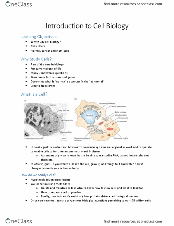
Types of microscopy: bright-field microscopy, phase-contrast microscopy, differential interference contrast microscopy, fluorescence microscopy, gfp and fluorescent fusion protein, confocal microscopy, deconvolution, fret, electron microscopy. Obtaining contrast in light microscopy by exploiting changes in the. Immunofluorescence microscopy (dead cells: antibodies are prepared for specific proteins (e. g. tubulin, actin, a sample cell culture is fixed on a slide in a process which kills and. Electron microscopy: electron microscopy can provide much better resolution than light and fluorescence microscopy, electrons have wavelike properties; wavelength of essentially. Lecture 3: isolation and analysis of cell organelles and molecules. Hoescht stain can penetrate into the nucleus and bind to. Darker bands on the developed x-ray film represent large concentrations of the protein of interest. Positions of marker proteins on the film need to be manually labeled with a pen. Lecture 4: protein synthesis and transport vinny aggarwal.