Physiology 2130 Study Guide - Final Guide: Bronchus, Thoracic Cavity, Angiotensin

10 Mar 2018
School
Department
Course
Professor
Document Summary
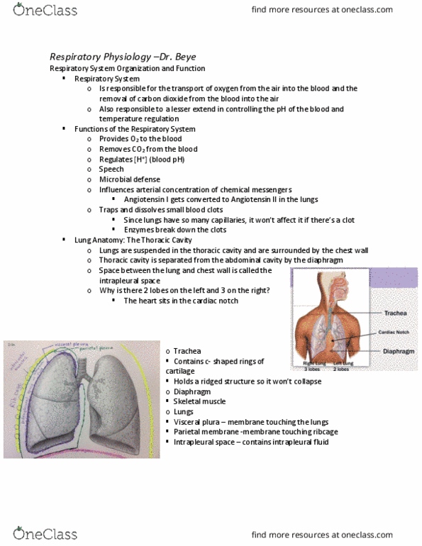
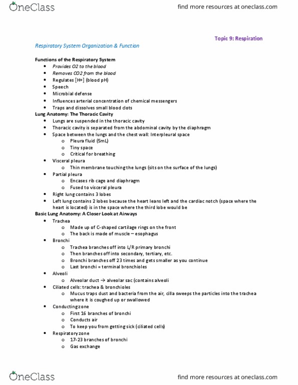
Thoracic cavity: the area above the diaphragm and between the ribcage. Trachea: airway made of c-shaped rings of cartilage in the front and muscle in the back. Diaphragm: dome-shaped skeletal muscle that separates the thoracic and abdominal cavity. Lungs: right has 3 lobes and left has 2 lobes: bottom of heart sits in cardiac notch of the left lung, each lung sits in its own double-layered sac. Intrapleural space: <1mm thick space between the pleura: filled with intrapleural fluid that allows the lungs to slide easily close to the chest wall. Intercostal muscles: between each rib to help us breathe. Primary bronchi: first set of branches off trachea: we have 2 left and right. Secondary bronchi: split of primary bronchus into 2 smaller tubes. Tertiary bronchi: split of secondary bronchus into 2 smaller tubes. Terminal bronchioles: the last set of bronchioles in the conducting zone. Respiratory bronchioles: part of the respiratory zone and splits multiple times into alveoli.