PSYC 4034 : Physiological Psychology Exam 4

Physiological Psychology Exam 4
CHAPTER 5
Visualizing the Brain
X-Ray
Works by passing a beam through tissue and you count on the tissue having different levels of radio
opacity. If the area isn’t opaque then the X ray will go right through and there will be no noticeable
difference.
Contrast X-Ray – procedures to artificially enhance the contrast of tissue inside the skull
o Pneumoencephalography – (First contract x-ray), inject a small amount of air into the CSF and it
enters into the brain. It creates a difference in the contrast between the CSF and the tissue
Used to visualize the ventricular system and look for deterioration of the cortex or
deformation in the ventricular system.
o Cerebral angiography – inject a radio- opaque liquid into the blood stream and whatever the blood
is carried to when x-rayed you can see very clearly the vascular structure. Shows displacement of
the vascular system, places of hyper vascularization (tumors)
This is an advanced technique of Frontal Lobotomies (Moniz)
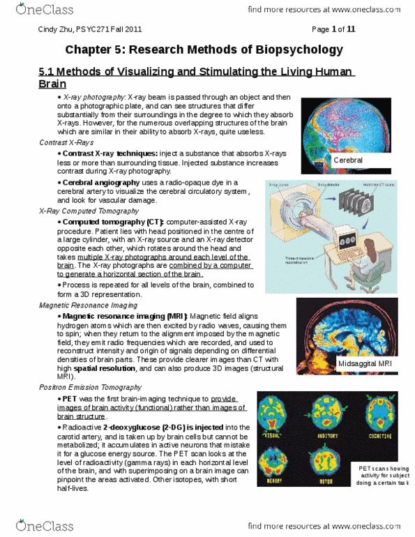
Computer tomography (CT scan) – refined x-ray technique. Difference is instead of using a plate,
photographs are done electronically and then the computer can enhance contrast artificially. It can detect
differences in contrast as it moves around the head. Gives a detailed picture of the brain.
o Drawback is you can only use this in the horizontal plane of the brain
o CT combined with angiography to visualize blood vessels. It can show the two major blood systems
that supply the head.
Magnetic Resonance Imagining (MRI)
The most detailed image of the living brain that we have.
They rely on the fact that atoms that make up the molecules of the brain can be made to emit a radio signal,
which is what the machine picks up.
o The atoms are polarized and they can align and orient themselves in the same direction. A radio
signal is taken up by the atoms and causes the electrons to get excited and move to a higher orbit.
The signal is turned off and they return to their original orbit, and they emit the energy as a radio
signal. Purpose of the magnet is so the atoms send their signals in the same direction.
o You can have sagittal plane views, frontal plane views, and horizontal plane views of the brain with
MRIs
Functional MRI or fMRI
o Best spatial and temporal resolution
o Only difference is the atom they target. Normal MRIs target hydrogen, fMRIs target oxygen. This is
because when the brain is active more oxygen is required in certain places (auto-regulation) and
you can observe metabolic rate, either at a base level or have the patient do something.
Diffusion Tensor Imaging (DTI)
Takes advantage of the fact that the movement of water molecules in bundles of white matter will not be
random, but are parallel to the axons that make up the bundles. This is an imaging method that uses a
modified MRI scanner to reveal bundles of myelinated axons in the human brain.
Positron emission tomography (PET) scan
Relies on a radioactive substance emitting detectable particles that can be pick up by machines, results in a
picture of the brain that’s not as detailed as an MRI, but has an advantage that you can look at what the
brain is doing. Usually used for metabolic studies.
Done by having a person take in a radioactive substance that is picked up by the blood and distributed by
the blood – commonly uses xenon (gas) and 2DG (2deoxyglucose). It’s a type of glucose that our cells do not
differentiate from other forms of glucose. 2DG is different from glucose because our cells can’t readily use
it, so it builds up in cells.
Disadvantage is that its slower than MRI; it takes time for the radioactive substance to disperse its way into
parts of the body
EEG
EEG stands for Electroencephalography
Electroencephalogram – the machine that records the activity of the brain
Nothing but a powerful amplifier. Detects the electrical signals from the head by macroelectrodes that are
attached to the scalp or put in a tight cap that the patient wears
o Macroelectrodes record any electrical activity that takes place immediately underneath them. This
electrical activity can include action potentials, post-synaptic potentials (EPSP, IPSI), electrical
impulses in the eyes, jaws, electrical activity that takes place in some of the cells in the blood
stream.
o Not a precise measurement of an individualized unit, but more generalized.
o the further away the signal generated is, the weaker it’s going to be by the time it gets to the
electrode
Allows us to triangulate the source of a signal and point out its origin.
o Seizure focus – very good for cortical and immediate subcortical foci for seizures
o General activity – measure of activity, used to determined EEG brain death, state of arousal, sleep
research
Microelectrodes – more precise EEG recordings; can record very discrete information from the brain, but is
invasive. Must be implanted into the brain.
o Stereotaxic Instrument – device used to implant microelectrodes. Normally a device that has a
circular array that’s bolted to your skull, and then they attach the electrodes
Allows you to precisely target a portion of the brain, but you are left to hope you implanted
the electrode in the correct location
Acts as a stereotaxic atlas and gives coordinates
Stereotaxic Surgery
Stereotaxic Surgery – implanting the tip of an electrode or cannula to a precise location in the depths of an
animal’s brain
Stereotaxic instrument (solid argument) is the ability to locate objects in space.
Stereotaxic Atlas
o The skill is composed of several bones that grow together and form sutures (seams). The heads of
newborn babies contain a soft spot at the junction of the coronal and sagittal sutures called the
fontanelle. Once the gap closes, the junction is called the Bregma
o The atlas contains photos that correspond to frontal sections taken at various distances, rostral and
caudal to the Bregma
o The Bregma Acts as a stereotaxic atlas and gives coordinates
Apparatus
o The device includes a head holder, which maintains the animal’s skull in the proper orientation;
holder for the electrode; calibrated mechanism that moved the electrode holder in measured
distance along three axis
Anterior to posterior
Dorsal to ventral
Lateral to medial
o Once the coordinates are obtained, the animal is anesthetized and placed in the apparatus and cut
the scalp open. The Bregma is located, a hole is drilled though the skull and the device lowered into
the brain by the correct amount.
Electron Microscopy
Used to see small anatomical structures
A beam of electrons is passed through the tissue to be examined
Scanning electron microscope provides less magnification, but provides 3D info about the shape and the
surface of a small object.
Anatomical Techniques
Histological techniques
After producing the lesion and observing its effects on behavior, we slice and stain the brain to observe it and see
the location of the lesion
Fixation and Sectioning
o Fixative (Fixing agent): a chemical such as formalin; use to prepare and preserve body tissue
o Formalin: the aqueous solution of formaldehyde gas, the most commonly used tissue fixative
Perfuse (Saline, then Formalin)
o “Sacrifice” the animals. Open the chest, expose the heart and insert a water and salt solution to
remove the blood. If you have blood in the capillary field it will pretty quickly begin to coagulate
and will block flow of the fixing agents
Remove the brain
o Chip off the skull, remove the brain and then store it in a Formalin solution to allow for the fixing
agent to get inside all of the brain tissue
Fix Brain to Formalin
o Formalin hardens it even more so that once it is completely fixed, it has the consistency of
something like an olive
o Prevents the tissue from being broken down by the catalytic enzymes that break apart protein that
cause autolysis; it prevents autolysis
o Kills bacteria
Section Brain
o Once the brain has been fixed, it is sliced into thin sections and stained to see various anatomical
details. Slicing is done with a microtome.
o 1. Used for brains or other sections of tissue that you separate. You make the brain harder by
embedding it into wax. Fixed brain into warm wax, the wax enters cells (ventricular system and all
of the cells), then let it cool and you have a really hard tissue to mount on a carrier arm. The
sections come off as ribbons that are covered with a transparent liquid known as a mounting
medium and placed on slides.
o 2. Freezing Microtome – When the liquid freezes, it doesn’t crystalize. Once on the microscope
slides, there is no differentiation between gray and white matter, it is very difficult to see where you
are in the brain, so the tissue must be stained
Staining the tissue
o Differential staining that will bind to certain parts of the tissue
o Myelin – the stain binds to the fat of myelin, called Myelin Stain
o Cell body – the stain binds to proteins that are in the cell body
Franz Nissl discovered that a dye, methylene blue would stain cell bodies of brain tissue.
Nissl substances consist of RNA, DNA and associated proteins located in the nucleus and
catered in the cytoplasm.
To verify the location of a brain lesion, a cell-body stain is used. Cell body stains also make it
possible to identify nuclear masses in the brain.
Most frequent dye used is cresyl violet.
The whole tissue looks purple and the stain is taken out of the lipids and left in the cell
body, resulting in a piece of tissue that allow you to look at nuclear group. This allows you
to analyze a single cell
o Membrane – Golgi Stain
If you take Golgi stain and apply it to neural tissue instead of just staining the myelin or
proteins, it stains all of the membrane of the cell and leaves you with the complete shape of
the neuron: The cell body and all of its neurites. It does the selectively by picking certain
neurons and staining them.
Central Nervous System Approaches
Brain lesions of subcortical regions are usually produced by passing electrical currents through a wire that
is coated with an insulting varnish, except for the tip.