BIOL 102 Chapter Notes - Chapter 6: Transmission Electron Microscopy, Scanning Electron Microscope, Endoplasmic Reticulum

2 Oct 2018
School
Department
Course
Professor
Document Summary
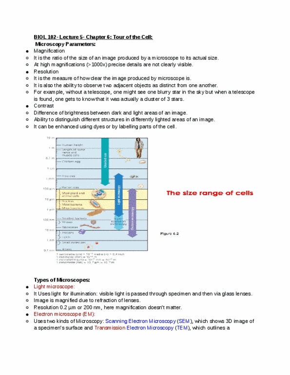
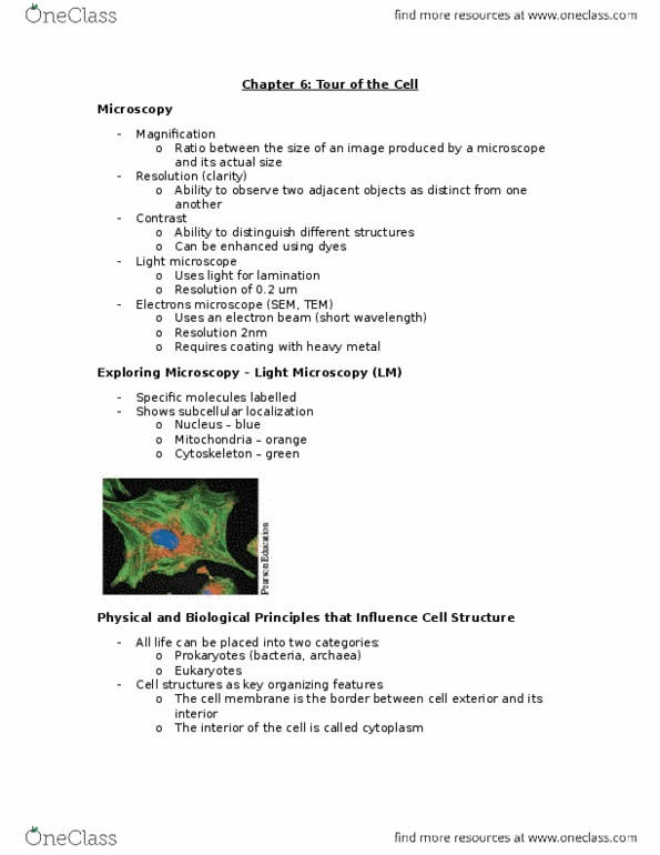
Light microscope- visible light is passed through the specimen and then through glass lenses: lenses refract the light to magnify the specimen, shows subcellular localization, nucleus is blue, mitochondria is orange, cytoskeleton is green. Magnification- ratio of a(cid:374) o(cid:271)je(cid:272)t"s size to its real size. Contrast- the difference in brightness between the light and dark areas of an image. Electron microscope- focuses a beam of electrons through a specimen or onto its surface, very good magnification. Scanning electron microscope- used for the detailed study of the topography (surface) of a specimen. Transmission electron microscope- to study the internal structure of cells. Dna is concentrated in a non-membrane-closed region called the nucleoid. Eukaryotic cells are more specialized and organised. Plasma membrane- phospholipid bilayer that encloses cytoplasm. Pili- allow cells to attach to other surfaces and each other. More organised, has membranes that partition the cell. Endoplasmic reticulum- membrane synthesis and other synthetic processes: rough has ri(cid:271)oso(cid:373)es, s(cid:373)ooth does(cid:374)"t.