PSY 4180 Chapter Notes - Chapter 7: Posterior Parietal Cortex, Transcranial Magnetic Stimulation, Two-Streams Hypothesis

5 Jun 2018
School
Department
Course
Professor

p. 360-372
Disorders of the Visual Pathway
The left half of each retina sends its projects to the right side of the brain, whereas the right half of each retina send its
projections to the left side of the brain.
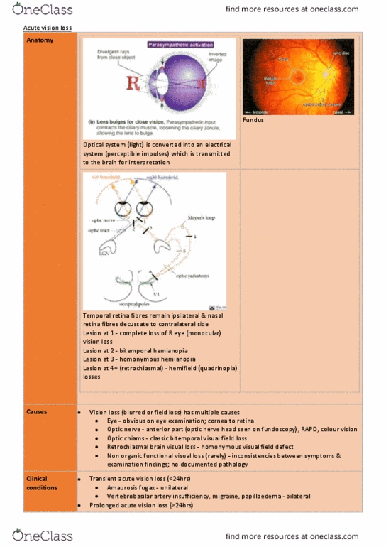
The representation of each side of the visual world seen by each eye is sent to the same place in area V1, and damage to V1
affects vision in both eyes.
Conversely, if a visual disturbance is restricted to just one eye, then the damage must be outside the brain, either in the retina
or in the optic nerve.
Different parts of the visual field are topographically represented in different parts of the area V1. Injury to a specific region
of V1 produces a loss of vision in a specific part of the visual world.
Destruction of the retina or option nerve of one eye produces monocular blindness, loss of vision in one eye.
A lesion of the medial region of the optic chasm severs the crossing fivers, producing bitemporal hemianopia, loss of vision of
both temporal fields.
A lesion of the lateral chasm result in a loss of vision of one nasal field, or nasal hemianopia.
Complete cuts of the optic tract, lateral genulate body, or area V1 result in (5) homonymous hemianopia — blindness of one
entire visual field.
• Some lesions are partial and result in quadrantanopia (loss of vision in one quarter of the visual field)
Small occipital lobe lesions often produce scotomas, small blind spots in the visual field. People are sometimes unaware of
this, because of nystagmus (constant tiny involuntary eye movements) and the brains ability to fill in information.
BK
Infarct — dead tissue. In the right occipital area, the size of a visual field detect is routinely measured with perimetry, a
standardized method in which the subject fixates on the black dot in a centre of a white screen and talks about when they
can see another light as it moves around on the screen.
Visual Noise — scintillating scotoma. Like a visual migraine
BK perceived locations of objects without being able to perceive content of objects, knowing you are seeing something in
your dead visual field but not actually being able to see it is known as blindsight.
DB: V1 Damage and Blindsight
• right calcarine fissure was removed surgically to exist an aginoma, a collection of abnormal blood vessels that
results in abnormal blood flow
• blindsight, but saying he is just guessing
• detect forms of movement, reports seeing something but isnt sure — again, a form of blindsight
• reports no conscious awareness of seeing but still is able to report on the movement and location of objects he can
not recognize
GY: V1 Damage and Conscious Vision
• using fMRI reveal that when he is aware of a moving stimulus projected to his blind field, activity occurs in V5 and
the prefrontal cortex in the hemisphere which is ipsilateral to the V1
• perhaps the V1 is not necessary for rudimentary visual awareness
• the prefrontal activity is presumably related to the brains attempt to understand the experience
Blindsight but movement — V5 +Frontal Lobe
find more resources at oneclass.com
find more resources at oneclass.com
Document Summary
The left half of each retina sends its projects to the right side of the brain, whereas the right half of each retina send its projections to the left side of the brain. The representation of each side of the visual world seen by each eye is sent to the same place in area v1, and damage to v1 affects vision in both eyes. Conversely, if a visual disturbance is restricted to just one eye, then the damage must be outside the brain, either in the retina or in the optic nerve. Different parts of the visual field are topographically represented in different parts of the area v1. Injury to a specific region of v1 produces a loss of vision in a specific part of the visual world. Destruction of the retina or option nerve of one eye produces monocular blindness, loss of vision in one eye.