BIO210Y5 Chapter Notes - Chapter 14: Superior Vena Cava, Pericardium, Aortic Valve

Document Summary
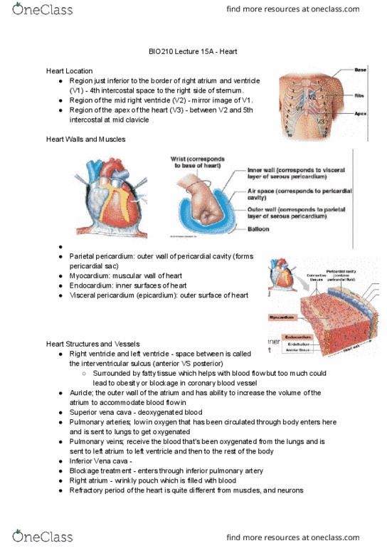
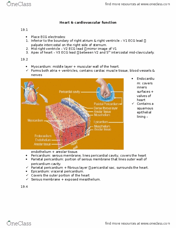
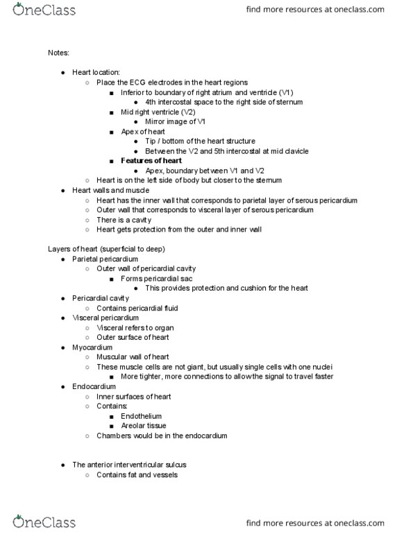
Region just inferior to the border of the right atrium and ventricle (v1) 4th intercostal space (space between ribs) to the right side of the sternum. Region of the mid right ventricle (v2) mirror image to v1. Region of the apex of the heart (v3) between v2 and the 5th intercostal at mid clavicle. Parietal pericardium: outer wall of pericardial cavity (forms pericardial sac) Visceral pericardium (epicardium): outer surface of the heart. Any inflammation of heart tissue it called carditis (endocarditis, pericarditis, cardiac tamponade) Auricle helps control volume changes; increase the capacity of the atrium, and so also increase the volume of blood that it is able to contain. The aorta is the largest artery in the body. The aorta begins at the top of the left ventricle, the heart"s muscular pumping chamber. The corresponding vein that carries deoxygenated blood from the upper half of the body is the superior vena cava.