BIO153H5 Chapter : 2014S_BIO153H5S_Lab1_Microscopy.pdf

Document Summary
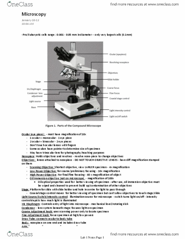
Be able to describe: the proper techniques for use of the compound and dissecting microscopes the proper use of the compound and dissecting microscopes total magnification of a compound microscope. Magnification, working distance, parfocal, parcentric, illumination, depth of field. How to prepare a wet mount the differences between the compound and dissecting microscopes. Be able to identify: the parts of the compound and dissecting microscopes and note their functions. The microscope remains a pivotal tool in learning and research in biology to this day. Typical prokaryotic or eukaryotic cells range from 0. 001 mm to 0. 05 mm in diameter, with only the very largest cells (0. 1 mm) entering into the range discernible by the human eye. Thus, microscopes are needed for detecting the existence of most cells and for visualizing cellular components. You will be using the compound light microscope and the dissecting microscope (also called the stereomicroscope).