Anatomy and Cell Biology 3319 Chapter Notes -Superior Sagittal Sinus, Dural Venous Sinuses, Dura Mater

20 Nov 2013
School
Department
Professor
Document Summary
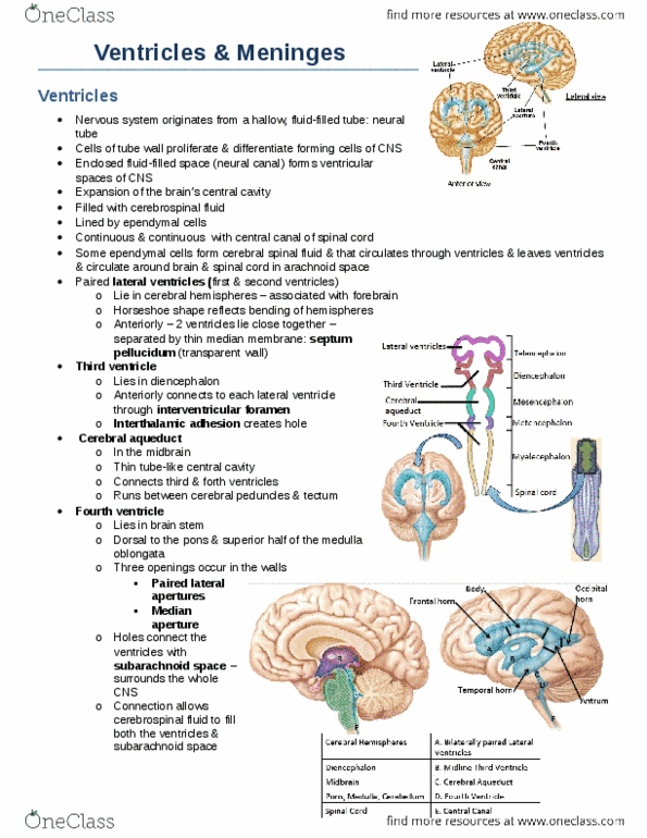
Ventricles are expansions of the brain"s central cavity, filled with csf and lined by epindymal cells. They are continuous with one another and the central canal of the spinal cord. Have paired lateral ventricles that lie in the cerebral hemispheres: horseshoe shape. Third ventricle lies in the diencephalon: anteriorly it connects to each lateral ventricle through an interventricular foramen. Fourth ventricle lies in the brain stem: dorsal to the pons, superior half of the medulla oblongata, caudally connects to the central canal, 3 openings occur in the walls: The holes connect the ventricles to subarachnoid space allows csf to fill both the ventricles and subarachnoid space. Blood-brain barrier: protects brain from harmful substances in the blood. These sinuses collect blood from the brain and conduct it to the large internal jugular veins of the neck. Largest dural sinus is the superior sagittal sinus in the superior midline.